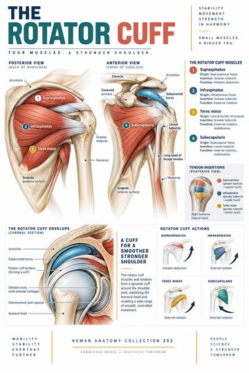
The rotator cuff is a group of four muscles and their tendons that connect the scapula to the proximal humerus. Together, they help stabilize the glenohumeral joint and contribute to arm movement. For medical educators and clinical teams, the cuff provides a useful way to explain how shoulder mobility depends on coordinated muscle activity as well as the shape of the joint.
The four rotator cuff muscles
The mnemonic SITS names the four muscles: supraspinatus, infraspinatus, teres minor and subscapularis. Each begins on the scapula, but their positions and principal actions differ.
| Muscle | Position on the scapula | Main contribution | Humeral attachment |
|---|---|---|---|
| Supraspinatus | Supraspinous fossa, above the scapular spine | Initiates abduction and supports the humeral head | Greater tubercle |
| Infraspinatus | Infraspinous fossa, below the scapular spine | Lateral rotation | Greater tubercle |
| Teres minor | Lateral border of the scapula | Lateral rotation; assists adduction | Greater tubercle |
| Subscapularis | Subscapular fossa on the anterior surface | Medial rotation; assists adduction | Lesser tubercle |
These descriptions identify principal actions; the contribution of an individual muscle varies with joint position and the task being performed. The teres major, despite its similar name, is not a rotator cuff muscle.
How the cuff supports the shoulder
The glenohumeral joint is formed by the head of the humerus and the glenoid cavity of the scapula. Its mobility makes dynamic muscular support particularly useful. Cuff tendons blend with the joint capsule and help keep the humeral head centered against the glenoid as larger muscles move the arm. The cuff works alongside the deltoid and muscles controlling the scapula; it does not stabilize or move the shoulder in isolation.
A useful teaching sequence is to first identify the scapular spine, then locate the supraspinatus above it and the infraspinatus below it. Find teres minor along the lateral scapular border. Finally, turn the scapula to its anterior surface to locate the subscapularis. This last step matters because a single posterior view cannot show all four muscles clearly.
Tendons and surrounding structures
The rotator cuff tendons approach the proximal humerus and reinforce the shoulder joint capsule. The supraspinatus tendon passes beneath the acromion. The subacromial bursa helps reduce friction between structures in this region. Nearby, the tendon of the long head of the biceps has a close anatomical relationship to the shoulder, but the biceps itself is not one of the four rotator cuff muscles.
When using a diagram in teaching, distinguish the muscle belly from its tendon and identify whether the illustration is an anterior, posterior or superior view. A simplified poster can support orientation, while a detailed atlas or other validated teaching resource can be used when exact attachment boundaries or anatomical variants matter.
Why this anatomy matters in practice
Rotator cuff tendons can be affected by injury or degenerative change. A tear can involve part or all of a tendon’s thickness, and more than one tendon may be involved. Symptoms and function vary; a diagram alone cannot establish the cause of shoulder pain or the appropriate treatment. In clinical education, identifying the involved structure and explaining its usual action can help patients understand the terminology used in an assessment or imaging report.
For an examination or clinical decision, use current professional guidance and the individual patient’s history, examination and relevant investigations. This overview is intended for education, not diagnosis or treatment.
Quick review
- Which cuff muscle is on the anterior surface of the scapula? Subscapularis.
- Which muscles principally rotate the humerus laterally? Infraspinatus and teres minor.
- Which cuff muscle helps initiate abduction? Supraspinatus.
- Where do the tendons attach? The greater or lesser tubercle of the humerus.
Putting the movements into context
Abduction takes the arm away from the side of the body in the anatomical plane; rotation turns the humerus around its long axis. These terms describe motion of the arm at the shoulder, not movement of the forearm alone. When discussing rotation, it helps to state the position of the elbow and shoulder, because the position changes how a movement is observed and how muscles contribute.
In a teaching session, compare the position of each muscle on the scapula with its route toward the humerus. The supraspinatus passes superiorly, while the infraspinatus and teres minor lie posteriorly. The subscapularis occupies the anterior surface. This spatial arrangement explains why a single view is incomplete and why a labeled anterior and posterior comparison can help learners build a three-dimensional picture.