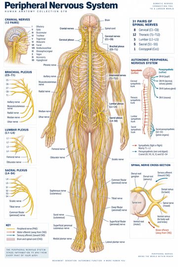
The twelve pairs of cranial nerves connect the brain with sensory structures, muscles and glands. Most are associated with the brainstem; the olfactory and optic nerves connect with forebrain structures. Roman numerals I–XII provide a consistent sequence for study and communication. The familiar sensory, motor or mixed labels summarize a nerve's fiber types; they do not replace an understanding of its individual branches and functions.
The twelve nerves at a glance
| Number | Nerve | Selected functions |
|---|---|---|
| I | Olfactory | Smell |
| II | Optic | Vision |
| III | Oculomotor | Most extraocular movements, eyelid elevation and parasympathetic pupil constriction |
| IV | Trochlear | Motor supply to superior oblique |
| V | Trigeminal | Facial sensation and muscles of mastication |
| VI | Abducens | Motor supply to lateral rectus |
| VII | Facial | Facial expression, taste from the anterior two-thirds of the tongue, and contributions to tear and salivary secretion |
| VIII | Vestibulocochlear | Hearing and balance |
| IX | Glossopharyngeal | Taste and sensation from the posterior tongue; roles in swallowing and salivation |
| X | Vagus | Pharyngeal and laryngeal functions, visceral sensation and parasympathetic output to thoracic and abdominal organs |
| XI | Accessory | Motor supply to sternocleidomastoid and trapezius |
| XII | Hypoglossal | Motor supply to most tongue muscles |
The table is a starting point rather than a complete inventory. For example, cranial nerve V has ophthalmic, maxillary and mandibular divisions; only the mandibular division carries its motor component to muscles of mastication. The vagus extends beyond the head and neck, so a head diagram alone cannot represent all of its targets.
Sensory, motor and mixed pathways
Olfactory, optic and vestibulocochlear are conventionally listed as predominantly sensory. Oculomotor, trochlear, abducens, accessory and hypoglossal are conventionally grouped as motor. Trigeminal, facial, glossopharyngeal and vagus are mixed. Some motor nerves also carry parasympathetic fibers, and a broad classification can hide important anatomical distinctions.
A practical way to study is to pair the name with a target or a question: smell for I, vision for II, eye movements for III, IV and VI, facial sensation for V, facial expression for VII, and hearing and balance for VIII. Then connect IX and X with the pharynx and swallowing, XI with neck and shoulder movement, and XII with tongue movement. These associations support memory but should not be used as substitutes for a complete neurological examination.
Reading a labeled diagram
Start by identifying the view. An inferior view of the brain helps show where several nerves emerge, whereas a lateral head view follows selected pathways toward their targets. The apparent origin of a nerve is not the same as tracing every nucleus, ganglion and branch. Because structures overlap, illustrations commonly separate or simplify them. Use the diagram to orient yourself, then consult a detailed atlas for foramina and pathway relationships.
Clinical context
Cranial nerve findings can help localize a neurological problem, but a single symptom rarely identifies a lesion by itself. Examination findings must be interpreted alongside onset, associated features and the rest of the neurological assessment. A mnemonic or poster cannot establish a diagnosis. For clinical decisions, consult current guidance and the relevant local pathway.
Quick review
- Which nerve carries vision? Optic, II.
- Which nerve supplies lateral rectus? Abducens, VI.
- Which nerve has three major sensory divisions in the face? Trigeminal, V.
- Which nerve carries hearing and balance information? Vestibulocochlear, VIII.