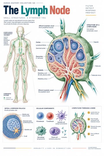
Lymph nodes are small, encapsulated organs located along lymphatic vessels. They filter lymph and provide organized spaces where immune cells can encounter material carried from tissues. Understanding the path through a node makes the relationship between lymphatic drainage and immune surveillance easier to explain in a classroom or consultation room.
Where lymph nodes fit in the lymphatic system
Some fluid leaving blood capillaries enters the interstitial spaces around cells. Lymphatic capillaries collect a portion of this fluid; once inside lymphatic vessels it is called lymph. Vessels carry lymph through lymph nodes and onward to larger lymphatic ducts, which return it to the venous circulation. Lymph does not travel through every node in the body: drainage follows regional pathways, and anatomical arrangements vary.
Nodes occur in groups, including cervical, axillary and inguinal regions, as well as within the thorax and abdomen. Their locations matter because lymph from a particular region commonly reaches particular groups first. This is a framework for understanding drainage, not a guarantee that any single finding identifies a cause.
Capsule, cortex and medulla
A connective-tissue capsule surrounds the node. Extensions of the capsule, called trabeculae, pass inward. Beneath the capsule is a network of sinuses through which lymph travels. The outer cortex contains lymphoid follicles, in which B lymphocytes are prominent. Some follicles develop germinal centers during immune responses.
The paracortex, deeper than the outer cortex, is relatively rich in T lymphocytes. It also contains high endothelial venules, specialized blood vessels through which many lymphocytes enter the node from the bloodstream. The inner medulla contains medullary cords and sinuses. Cords contain immune cells, including plasma cells; sinuses are channels through which lymph moves toward the exit.
These regions communicate continuously. A diagram may use distinct colors to make zones easy to recognize, but the boundaries are not rigid walls separating immune cells completely.
Follow the lymph through a node
Multiple afferent lymphatic vessels typically bring lymph into the convex surface of a node. Lymph passes into the subcapsular sinus and then through cortical and medullary sinuses. It leaves through one or more efferent lymphatic vessels at the hilum, the indented region where blood vessels also enter or leave.
Macrophages and other immune cells in the node interact with material carried by lymph. The architecture slows and channels flow so immune surveillance can occur. Not all immune responses begin in lymph nodes, and a node is not simply a passive sieve: it is living tissue with circulating and resident immune cells.
Lymph nodes versus the spleen
Both are secondary lymphoid organs, but their principal input differs. Lymph nodes sample lymph arriving from tissues; the spleen filters blood. Confusing these routes can lead to misleading explanations of the lymphatic system. The thymus and bone marrow have different roles in lymphocyte development and should also be distinguished from the nodes.
Clinical context and careful communication
A node may enlarge for several reasons, including immune responses, infection, inflammatory conditions or malignancy. Its location and other characteristics can inform assessment, but a diagram or the presence of enlargement alone does not establish a diagnosis. Clinical interpretation depends on the full history, examination and investigations when appropriate.
For patient education, describe a node as an immune checkpoint along lymphatic drainage pathways, then clarify that nodes are normal structures throughout the body. If discussing a specific finding, use the treating clinician's explanation and avoid suggesting that a poster replaces individualized assessment.
How to read a lymph-node diagram
Find the capsule and convex surface first. Trace arrows from afferent vessels into the subcapsular sinus, across the node and out at the hilum. Then identify cortex, paracortex and medulla. Check whether the drawing also labels a blood vessel: blood circulation and lymphatic flow are related but distinct pathways. Orientation and the number of vessels are simplified in many illustrations.
Quick review
- What enters through afferent lymphatic vessels? Lymph from upstream tissues and vessels.
- Where does lymph leave the node? Through efferent lymphatic vessels at the hilum.
- Which organ filters blood rather than lymph? The spleen.
- Where are many lymphoid follicles? In the outer cortex.
Why regional drainage matters
Lymphatic vessels connect tissue regions to chains of nodes, and efferent vessels may lead to additional nodes before lymph reaches a larger collecting trunk. The arrangement is not identical in every person. When using a diagram to teach regional anatomy, label the territory and direction of flow and distinguish a general pathway from a map of an individual patient. Surgical or inflammatory changes can alter normal drainage patterns.