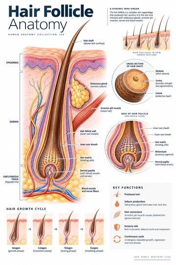
A hair follicle is an epithelial structure that extends from the epidermis into the skin and produces a hair. It is associated with a sebaceous gland, sensory nerve endings and, in many locations, an arrector pili muscle. Together these structures illustrate how the surface of the skin connects with deeper tissues.
Hair shaft and root
The visible hair shaft projects above the skin surface. The root lies within the follicle, below the surface. At its deeper end, the expanded hair bulb surrounds a connective-tissue projection called the dermal papilla. Cells in the hair matrix divide and contribute to the growing hair; melanocytes associated with the matrix contribute pigment to the hair.
A hair is commonly described in terms of a cuticle, cortex and, in some hairs, a central medulla. The exact appearance differs with hair type. A diagram enlarged for teaching makes the layers visible but does not represent every follicle at the same stage of its growth cycle.
The follicle and its surrounding tissues
The follicle is an invagination of epithelium that extends into the dermis and can reach deeper tissue depending on location and growth stage. Epithelial root sheaths surround the hair root. Connective tissue, blood vessels and nerves around the follicle support and interact with it. The dermal papilla contains vessels that supply the growing region indirectly; the portion of hair projecting beyond the skin is not itself living tissue.
Skin proper consists of the epidermis and dermis. The hypodermis or subcutaneous tissue lies beneath them. Some teaching graphics group the hypodermis with the skin for orientation, so it helps to state which definition is being used.
Sebaceous gland and arrector pili
A sebaceous gland usually empties oily sebum into the upper portion of a hair follicle. Its secretion helps condition the hair and skin surface. Sebaceous glands are associated with many follicles, although distribution and size vary by body site. Not every gland is best explained through a single idealized hair diagram.
The arrector pili is a small band of smooth muscle connected to the follicle and surrounding dermis. When it contracts, the hair can become more upright and the skin surface may pucker, producing goosebumps. Sympathetic activation can cause this response. It is a recognizable example of the connection between skin appendages, smooth muscle and autonomic control.
Hair growth is cyclical
Follicles cycle through growth, transition and rest phases, commonly called anagen, catagen and telogen. A hair may subsequently shed and a new cycle begin. Neighboring follicles are not all synchronized, and the duration of a phase differs by body site and individual. A static illustration cannot show the whole cycle; compare several labeled stages when explaining growth or shedding.
The follicle contains populations of cells that support renewal. Growth and pigmentation change with age and are influenced by genetics, hormones, nutrition, illness and other factors. These relationships are complex and should not be reduced to a single cause in patient education.
Reading a cross-section
Locate the epidermal surface and follow the hair shaft downward to the root and bulb. Identify the dermal papilla at the base, then look for the sebaceous gland opening near the upper follicle. Trace the arrector pili from the follicle toward the dermis. If a sensory nerve plexus is shown, distinguish it from blood vessels; both can appear as thin branching lines in a simplified diagram.
A follicle may be cut obliquely in a histological section, making its structures look different from a neat lengthwise poster. The appearance also depends on the growth phase. Use the illustration as an orientation aid and a histology reference for detailed identification.
Clinical context
Changes in hair growth or shedding may have many causes. Location, timing, associated skin changes, medications and medical history can all matter during assessment. An illustration cannot diagnose an individual condition or determine treatment. In education, the follicle diagram helps explain which structures are being discussed and why a change at the root differs from damage to an exposed hair shaft.
Quick review
- What is visible above the skin? The hair shaft.
- What surrounds the root? The follicle and its root sheaths.
- Where does a sebaceous gland commonly open? Into the follicle.
- What causes a hair to stand more upright? Contraction of the arrector pili muscle.
Variation across body sites
Follicles differ in size, depth, pigmentation and the kind of hair they produce. The relationship between follicle and sebaceous gland is easier to see in a diagram of a typical pilosebaceous unit than in every real section. Hair-bearing skin also differs from the thick skin of palms and soles, which lacks hair follicles. These differences make the location of a sample important when interpreting a diagram or histological section.