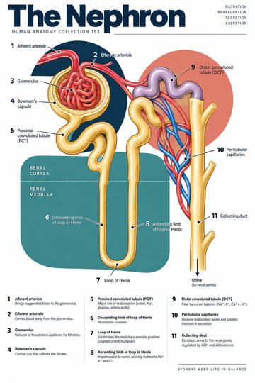
The nephron is the kidney's microscopic functional unit. Its renal corpuscle produces an initial filtrate from blood, and its tubular segments modify that fluid. Following the path from glomerulus to collecting duct helps explain how the kidneys regulate water, electrolytes and the excretion of waste.
The renal corpuscle
The renal corpuscle consists of the glomerulus, a tuft of capillaries, and the surrounding glomerular or Bowman's capsule. Blood enters the glomerulus through an afferent arteriole and leaves through an efferent arteriole. Water and small solutes pass across the filtration barrier into the capsular space. Blood cells and most large plasma proteins normally remain in the circulation.
Filtration is selective rather than a literal sieve with a single layer. Fenestrated capillary endothelium, the glomerular basement membrane and filtration slits between podocyte processes contribute to the barrier. A diagram of the corpuscle is schematic and cannot show all microscopic details at the same scale.
Proximal tubule
Filtrate enters the proximal convoluted tubule. Here substantial amounts of filtered water and solutes, including sodium, glucose and amino acids, are reabsorbed under normal conditions. Tubular secretion also moves selected substances from blood into tubular fluid. Reabsorption means movement from the tubule back toward the blood; secretion means movement in the opposite direction into the tubule. The terms describe direction, not whether a substance is useful or harmful.
Loop of Henle
The nephron loop descends into the medulla and returns toward the cortex. Segment permeability and transport differ. In the simplified teaching model, the thin descending limb is permeable to water, while the thick ascending limb reabsorbs salts and is relatively impermeable to water. These differences help establish the medullary osmotic gradient, which contributes to the kidney's ability to concentrate urine. Actual segment characteristics vary and should not be flattened into a claim that every descending or ascending segment behaves identically.
Distal tubule and collecting system
The distal convoluted tubule continues adjustment of solute handling. Fluid then enters connecting segments and collecting ducts. Collecting ducts receive fluid from multiple nephrons and are usually described separately from the nephron proper, although diagrams often show them together. Antidiuretic hormone can increase water permeability in appropriate collecting-duct cells, while aldosterone influences sodium reabsorption and potassium secretion in the distal nephron and collecting system.
The final composition of urine reflects filtration, reabsorption and secretion across multiple segments. Fluid leaving collecting ducts passes through the renal papillae into calyces and then the renal pelvis; it is not already outside the kidney at the end of the nephron loop.
Blood supply and tubule relationships
The efferent arteriole gives rise to peritubular capillaries, and in juxtamedullary nephrons vessels called vasa recta run into the medulla. These blood vessels participate in exchange with surrounding tissue. Trace blood and tubular fluid separately on a poster: blood does not flow along the tubular lumen, and filtrate does not return to the bloodstream without crossing an epithelial and interstitial route.
Clinical and teaching context
Kidney function is assessed using the patient's history, examination and appropriate tests. Estimated glomerular filtration rate and urine findings provide different types of information; neither is a picture of a single nephron. Many conditions and medicines can affect renal physiology. This educational overview does not diagnose disease or provide individual treatment advice.
Quick review
- Where does filtration begin? At the glomerular capillaries and filtration barrier.
- What is reabsorption? Movement from tubular fluid toward the blood.
- What is secretion? Movement into tubular fluid.
- What is the role of the loop? Segment-specific transport helps establish the medullary gradient.