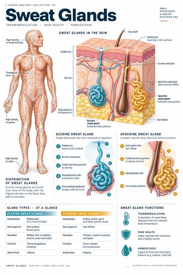
Sweat glands are coiled tubular glands associated with the skin. Eccrine and apocrine glands differ in distribution, ducts and secretion. A labeled skin section can help clinicians and educators explain how a gland deep in the skin connects to the surface or to a hair follicle.
Eccrine sweat glands
Eccrine glands are widely distributed across much of the body, with high concentrations in areas including the palms, soles and forehead. Their secretory portion is coiled in the dermis or deeper tissue. A duct carries secretion toward a pore on the skin surface. Myoepithelial cells around the secretory portion assist the movement of secretion into the duct.
Eccrine sweat is largely water with electrolytes. As sweat evaporates from the skin surface it carries heat away, supporting thermoregulation. The amount that evaporates depends on environmental conditions: fluid on the skin does not provide the same cooling if it cannot evaporate effectively. Sweating is one part of temperature regulation, which also involves blood flow and behavioral responses.
Apocrine sweat glands
Apocrine glands are concentrated in selected regions, including the axillae and anogenital area. Their ducts typically open into hair follicles. Their activity becomes more prominent around puberty, and their secretion contains organic substances. Skin bacteria can break down components of the secretion and contribute to body odor.
The word “apocrine” is a traditional gland classification; it should not be taken to mean that every aspect of the secretion mechanism is captured by the name. The glands differ from sebaceous glands, which produce sebum and commonly also empty into hair follicles. A hair-follicle diagram can show both structures, but their secretions and functions are not interchangeable.
Nervous control and temperature
Sweating is regulated by autonomic pathways. Most thermoregulatory eccrine glands are activated by sympathetic cholinergic fibers, an exception to the common introductory association of sympathetic postganglionic signaling with noradrenaline. Responses differ by body region and stimulus; emotional sweating can be prominent on the palms and soles. A single diagram cannot show all control pathways or individual variation.
Heat balance also depends on skin blood flow, air movement, humidity, clothing and hydration. Explaining sweat glands as one component of a wider regulatory system helps avoid treating a change in sweating as proof of a particular diagnosis.
Reading a skin diagram
Locate the epidermis, dermis and underlying subcutaneous tissue. Follow an eccrine duct from its coiled gland to the surface pore. Then find an apocrine gland and trace its duct toward a hair follicle. Structures are often enlarged and arranged neatly for teaching; real glands and ducts occupy three-dimensional space and may appear differently in a histological section.
Skin proper consists of epidermis and dermis. Many diagrams show the hypodermis beneath these layers for context. State the convention being used when describing “layers” so learners do not confuse the boundaries.
Clinical and teaching context
Sweating patterns vary with temperature, activity, medication and health conditions. Reduced or excessive sweating can have many causes, and symptoms should be interpreted in context. This guide and its accompanying image are educational aids, not a diagnostic tool or treatment recommendation.
For patient education, a concise explanation is that eccrine sweat reaches the skin surface and evaporation helps cool the body. A separate explanation of odor can describe the interaction between gland secretions and skin bacteria without suggesting that all sweat has the same composition.
Quick review
- Which glands are widespread and important for cooling? Eccrine glands.
- Where does an eccrine duct usually open? At the skin surface.
- Where does an apocrine duct typically open? Into a hair follicle.
- How does sweat cool the body? Primarily through evaporation from the skin.