The cardiac cycle describes the recurring mechanical events that move blood through the heart. A useful starting point is to distinguish systole, when a chamber contracts, from diastole, when it relaxes and fills. Atrial and ventricular phases overlap, so a diagram should specify which chamber is being described.
From ventricular filling to atrial contraction
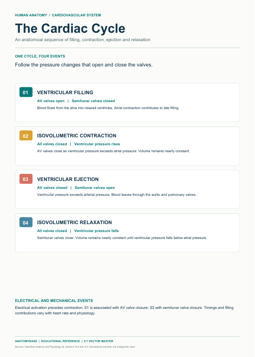
During much of ventricular diastole, ventricular pressure is below atrial pressure. The mitral and tricuspid valves are open, and blood flows from the atria into the ventricles. The aortic and pulmonary valves are closed. Filling is initially rapid and then slows; the slower interval is often called diastasis. Atrial systole adds blood to the ventricles near the end of diastole. The proportion contributed by atrial contraction varies with heart rate and ventricular properties.
Isovolumetric contraction
Ventricular electrical activation precedes mechanical contraction. As ventricular pressure rises above atrial pressure, the atrioventricular valves close. The first heart sound, S1, is associated with closure and the resulting vibrations. For a brief interval the semilunar valves are also closed, so ventricular volume remains nearly constant even though pressure rises. This interval is called isovolumetric contraction.
Ventricular ejection
When left ventricular pressure exceeds aortic pressure, the aortic valve opens; on the right, the pulmonary valve opens when right ventricular pressure exceeds pressure in the pulmonary trunk. Blood is ejected into the great arteries. Ejection is commonly described as rapid and then reduced. The two sides follow the same general sequence but operate at different pressures. Valve opening and closure reflect pressure differences rather than an independent muscular opening mechanism.
Relaxation and the next cycle
As the ventricles relax and their pressures fall below arterial pressures, the aortic and pulmonary valves close. Their closure contributes to the second heart sound, S2. The ventricles then relax briefly while both sets of valves are closed: isovolumetric relaxation. Once ventricular pressure falls below atrial pressure, the atrioventricular valves open and filling resumes.
A cycle can be entered at any point; what matters is the order and the pressure relationships. At faster heart rates, the length of the phases changes and diastole generally shortens proportionately more than systole. A teaching figure showing one cycle at one rate does not establish a universal duration for every patient.
Connecting pressure, volume and ECG
End-diastolic volume is the ventricular volume just before contraction; end-systolic volume is the volume remaining after ejection. Their difference is stroke volume. In an idealized diagram, pressure rises without a change in volume during isovolumetric contraction, and volume falls during ejection. The P wave represents atrial depolarization, the QRS complex ventricular depolarization, and the T wave ventricular repolarization. Electrical signals precede associated mechanical events; an ECG trace alone is not a direct pressure or valve recording.
Clinical and teaching context
Valve disease, changes in rhythm and altered ventricular filling can change the timing and relationships shown in a simplified cycle. A diagram supports discussion of normal physiology but cannot diagnose a murmur, arrhythmia or heart failure. Clinical interpretation requires the patient's context and appropriate examination and investigations.
For a patient-friendly explanation, describe valves as one-way structures responding to pressure: the heart fills, pressure rises, blood is pumped out, and the chambers relax for the next beat. Avoid implying that all filling occurs only after atrial contraction.
Quick review
- When are all four valves closed? During the idealized isovolumetric contraction and relaxation intervals.
- What opens the aortic valve? Left ventricular pressure exceeding aortic pressure.
- What does stroke volume describe? End-diastolic minus end-systolic ventricular volume.
- What does S2 relate to? Closure of the semilunar valves.