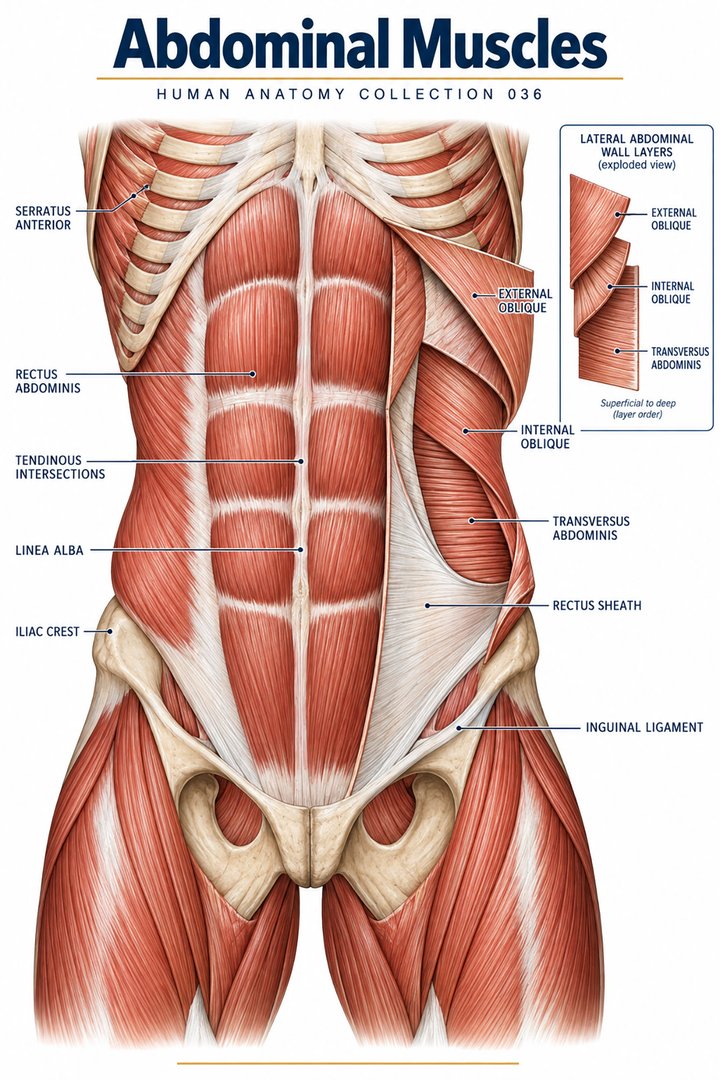
Abdominal Muscles
Individual poster · 300 DPI print files
Regular $9.90
Explore poster ↗$4.90USD
Offer price · Save $5.00Individual poster · 300 DPI print files
Reduced web preview. Separate high-resolution print files are supplied with the digital download.
ANATOMY 036
A modern anatomy illustration exploring abdominal muscles, designed for educational and decorative use.
Purchases are not available yet. Checkout and download delivery are being prepared.
Sold by Kunstolymp.de · Germany
Compare prices & the complete collection ↗This is a DIGITAL DOWNLOAD. No physical product will be shipped. After purchase, download your files and print them at home, at a local print shop, or through an online professional printing service.
The separate 300 DPI print files are ideal for 24×36-inch posters (approximately 60×90 cm). ISO and 5×7 formats are included. The white border lets you adjust the layout or trim to fit other frame sizes; check the preview before printing to keep the illustration and labels visible.
The planned bundle delivery includes a PDF information sheet with a link to the relevant Google Drive folder and download instructions. The store is currently in preview and purchases are not yet available.
A science-inspired choice for a student’s study corner, an educator’s classroom, or an anatomy enthusiast’s wall. Final licensing terms will apply.
CLINICAL CONTEXT
The abdominal wall is formed by rectus abdominis, external and internal obliques, transversus abdominis, fascia, and their attachments to ribs, pelvis, and thoracolumbar structures. These tissues support trunk movement, breathing, intra-abdominal pressure, coughing, continence, and load transfer. Their surface anatomy helps clinicians identify scars, asymmetry, rectus diastasis, masses, and hernias. However, pain in the abdominal wall can closely resemble intra-abdominal disease, and tenderness over a muscle never safely excludes appendicitis, cholecystitis, bowel obstruction, pancreatitis, urinary disease, pelvic pathology, ischaemia, or other urgent causes.
Acute strain can follow sudden twisting, heavy lifting, forceful coughing, sport, pregnancy-related change, or a new exercise programme. Patients may describe focal pain, spasm, pain on coughing or sit-up, bruising, and difficulty turning in bed. Chronic overload can affect athletes and manual workers. Hernia presents as a bulge or exertional discomfort and may be complicated by obstruction or strangulation. Diastasis recti is a widening of the linea alba and can affect function and body image, but it is distinct from a true fascial defect. Systemic symptoms, persistent vomiting, gastrointestinal bleeding, fever, weight loss, altered bowel function, urinary symptoms, pregnancy possibility, or pain that becomes severe or generalised require careful assessment.
History includes pain onset and migration, prior operations, trauma, pregnancy and menstrual history, anticoagulation, bowel and urinary symptoms, medication, and associated systemic features. Examination begins with vital signs and overall appearance, then inspection, cough impulse, palpation, percussion, and targeted assessment of peritonism, masses, spine, hips, and hernia sites. Urinalysis, pregnancy testing, blood tests, ultrasound, CT, or surgical and gynaecological input may be required. For uncomplicated strain, a plan often includes relative rest, analgesia tailored to risk, avoidance of painful overload, and progressive physiotherapy. Symptomatic or complicated hernias may require surgical evaluation. Underlying visceral or pelvic disease needs cause-specific treatment rather than an abdominal-wall exercise plan.
Guided breathing, graded core rehabilitation, yoga adapted to symptoms, and behavioural pain approaches can complement rehabilitation after serious pathology is excluded. They do not treat obstruction, infection, haemorrhage, or ischaemia. Avoid aggressive abdominal manipulation in acute or unexplained pain, after recent surgery, or where hernia or pregnancy complications are possible. Herbal and “detox” products can cause diarrhoea, liver injury, electrolyte disturbance, or interactions; patients should disclose them without judgment.
Seek urgent local medical care for severe or escalating abdominal pain, a rigid abdomen, persistent vomiting, blood in vomit or stool, fainting, fever with severe illness, a painful irreducible hernia, pregnancy-related pain or bleeding, or inability to pass stool or gas with distension.
Further reading: MedlinePlus: muscle strains · WHO: safe, evidence-informed integrative care
Explain the working diagnosis, important alternatives, planned tests, expected course, and specific safety-netting in language the patient can use. Invite questions about treatment goals, capacity, cost, culture, and complementary therapies. Use a professional interpreter when language access could affect consent or understanding, and confirm clear arrangements for timely clinical review in writing. Document the reasoning, reassess response to treatment, and coordinate care across professions when illness affects more than one system. Teaching anatomy is most useful when it supports this partnership: accurate localisation, respectful listening, timely escalation, and a plan that remains responsive to the individual patient.
HUMAN ANATOMY COLLECTION · 001—300
A closer look at what makes us human. Discover anatomy posters for your study space, classroom, and everyday curiosity.
DIGITAL DOWNLOADS · 300 DPI PRINT FILES
EXPLORE BY SYSTEM
LEARN & EXPLORE
Detailed English guides with Latin terminology, diagrams, real images, and clinical learning connections.
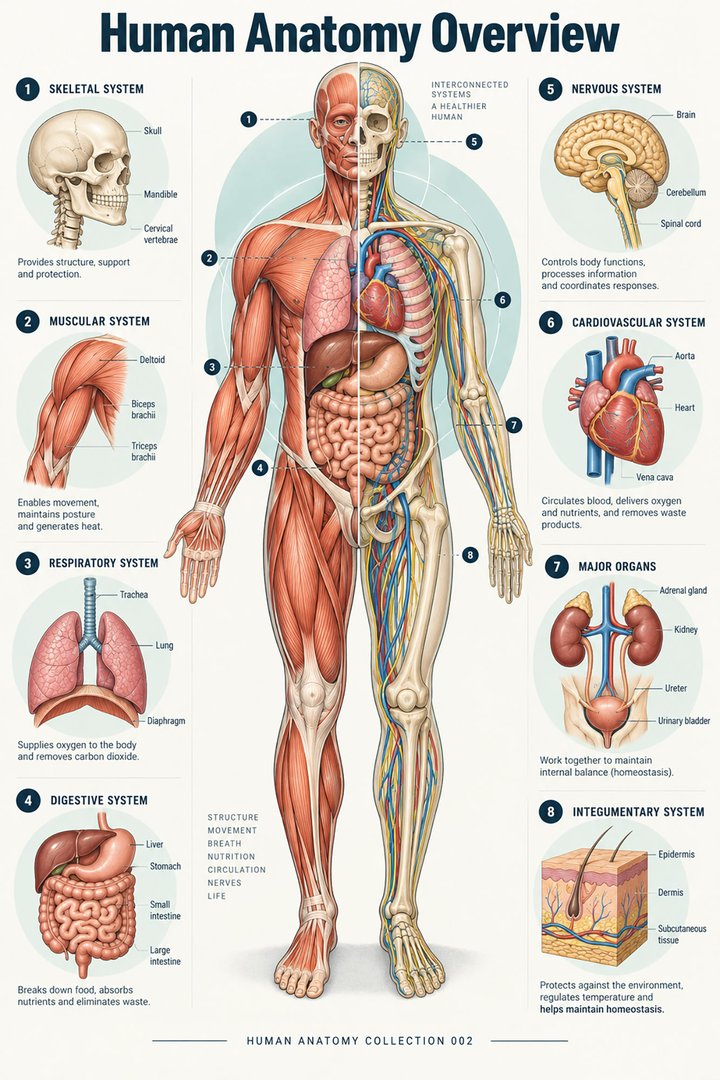 Human Anatomy Diagrams ↗
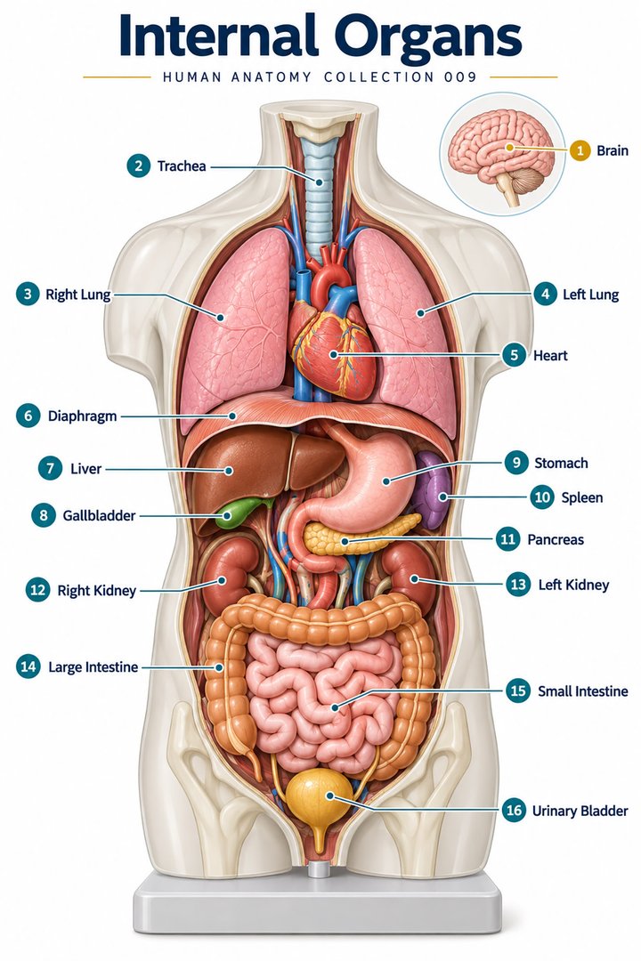 Human Body Organs ↗
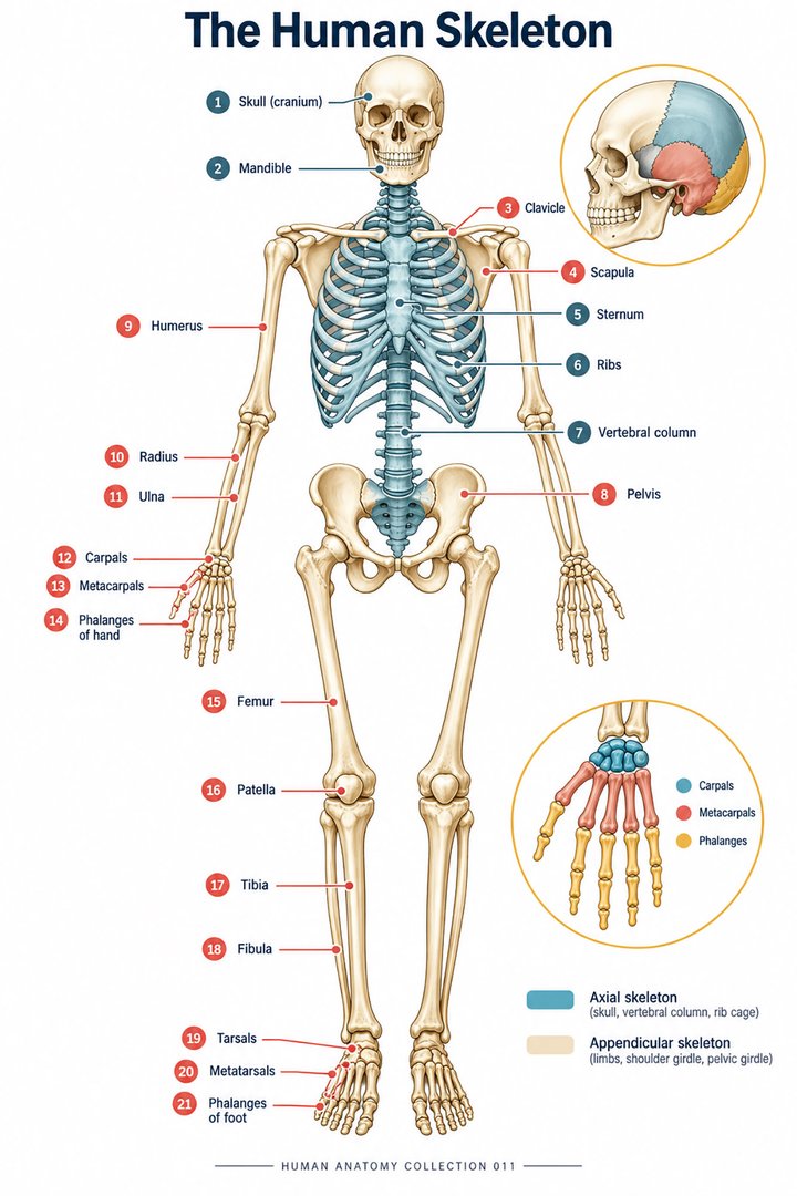 Skeletal System Anatomy ↗
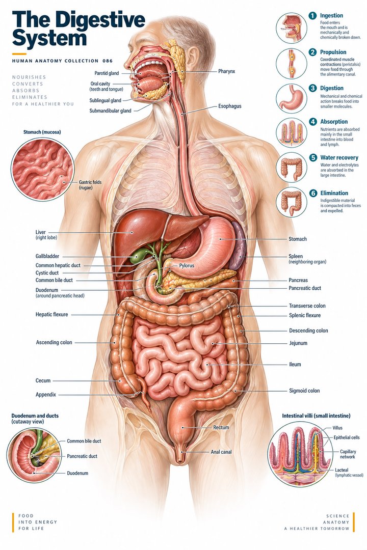 Human Digestive System ↗
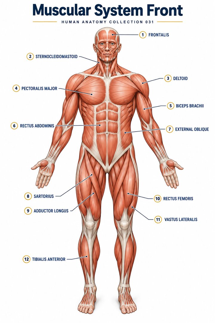 Muscular System Overview ↗
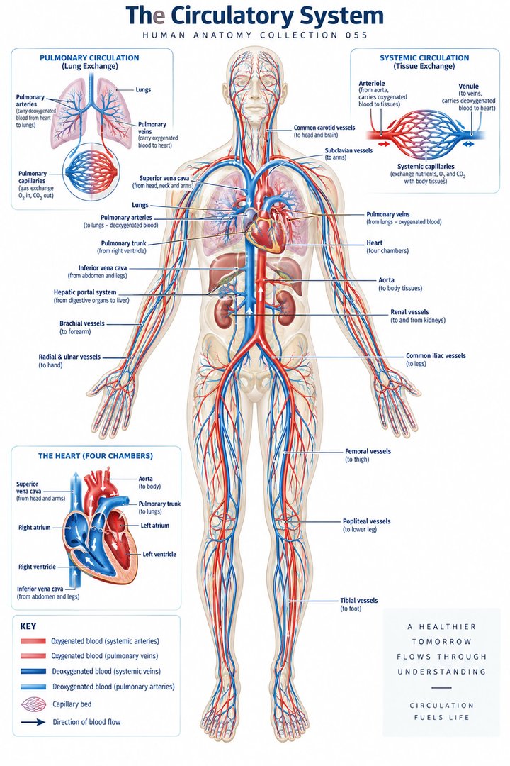 Human Circulatory System ↗
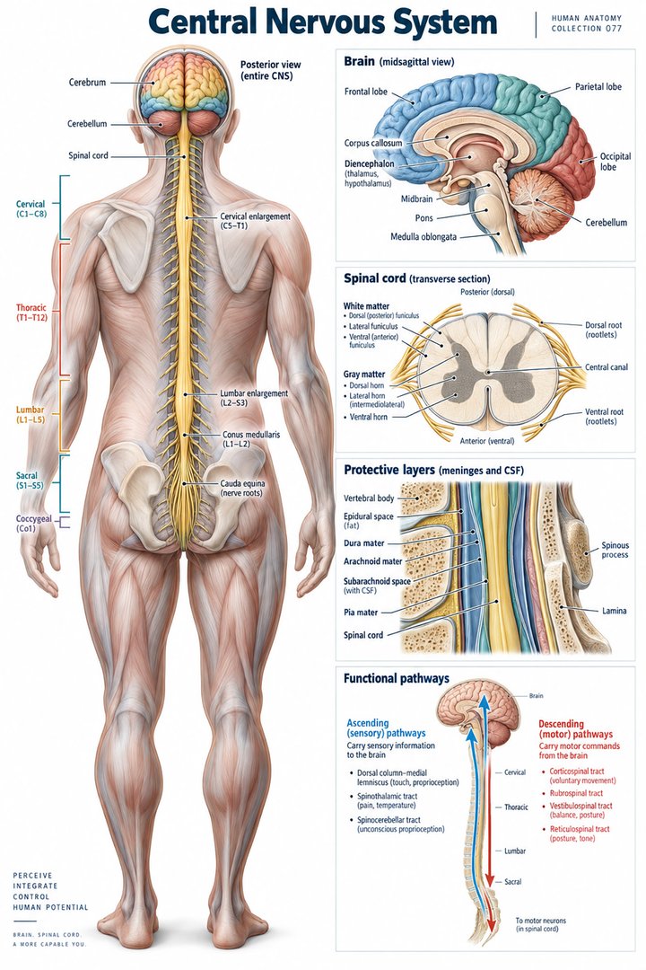 Human Nervous System ↗
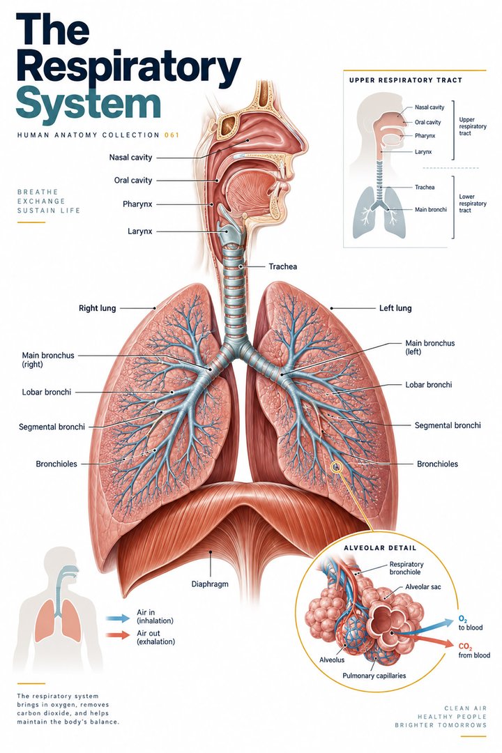 Respiratory System Functions ↗
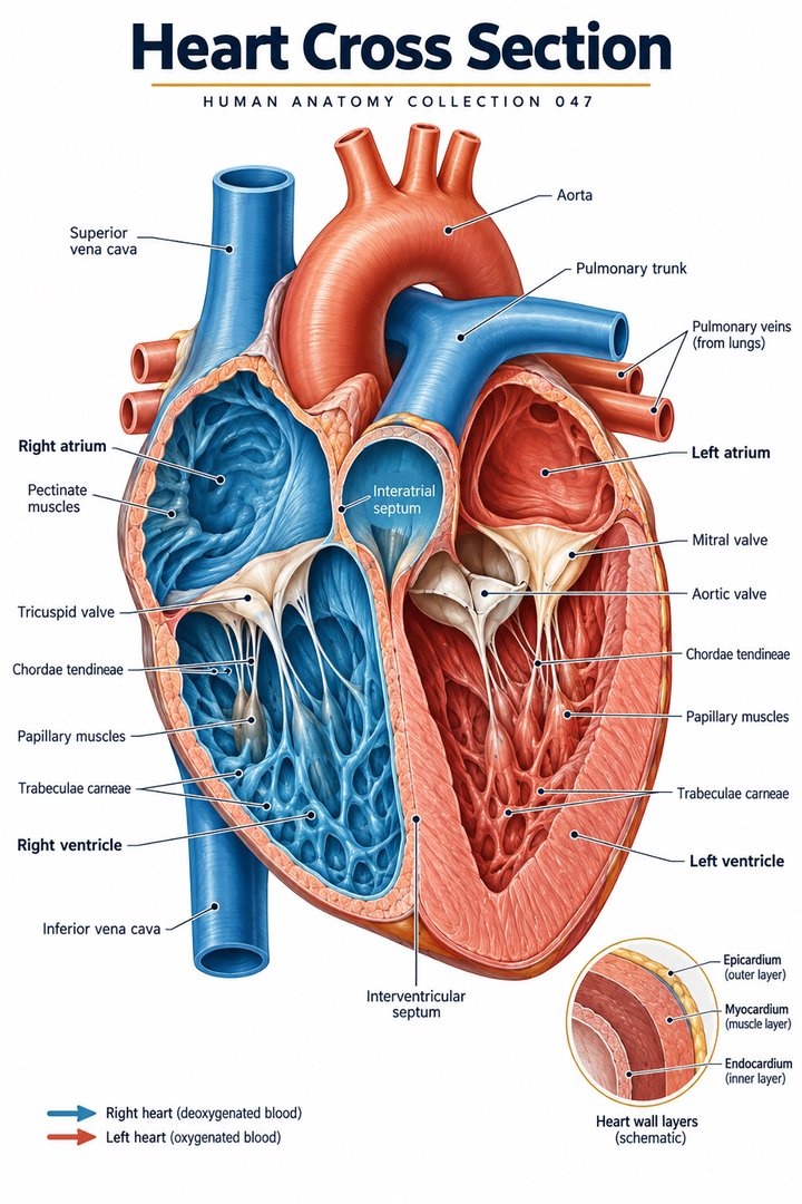 Human Heart Structure ↗
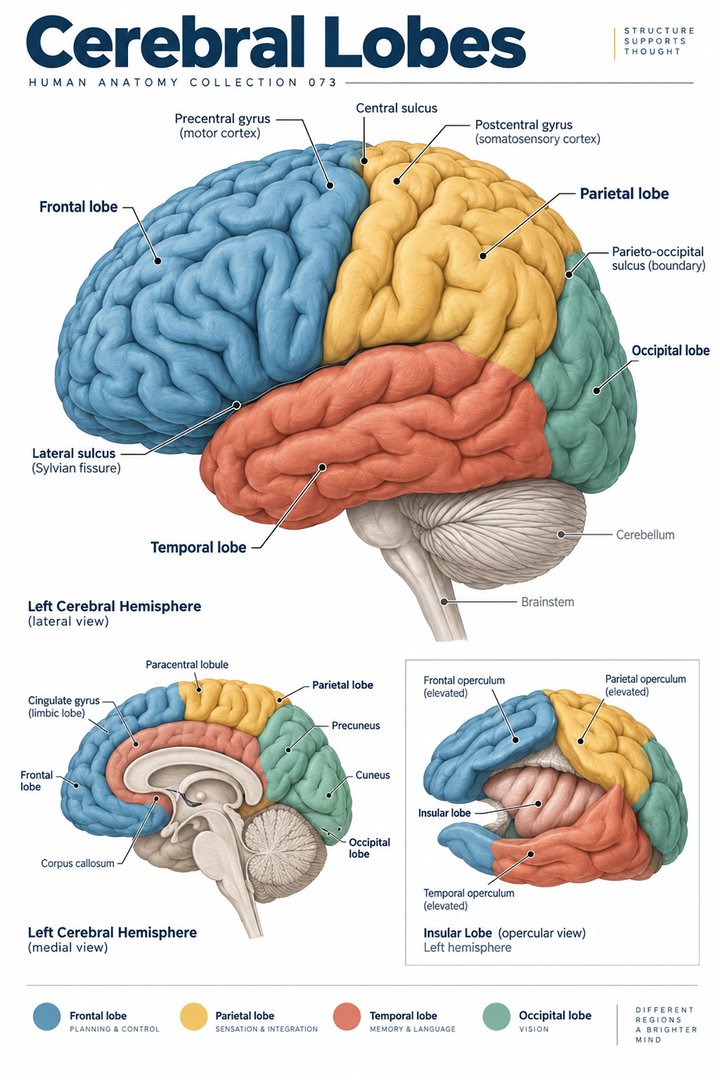 Human Brain Lobes ↗
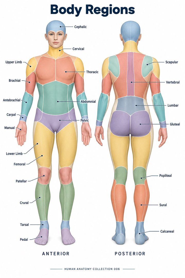 Human Body Parts ↗
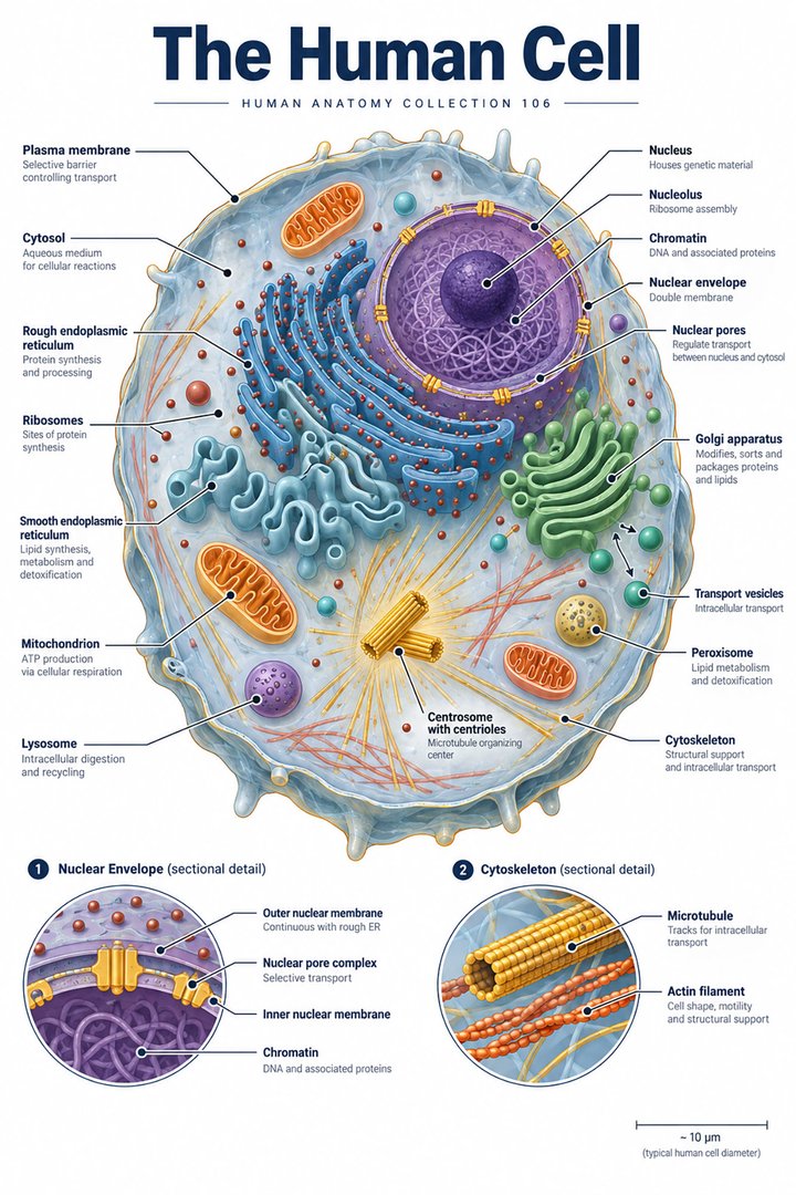 Human Cell Structure ↗
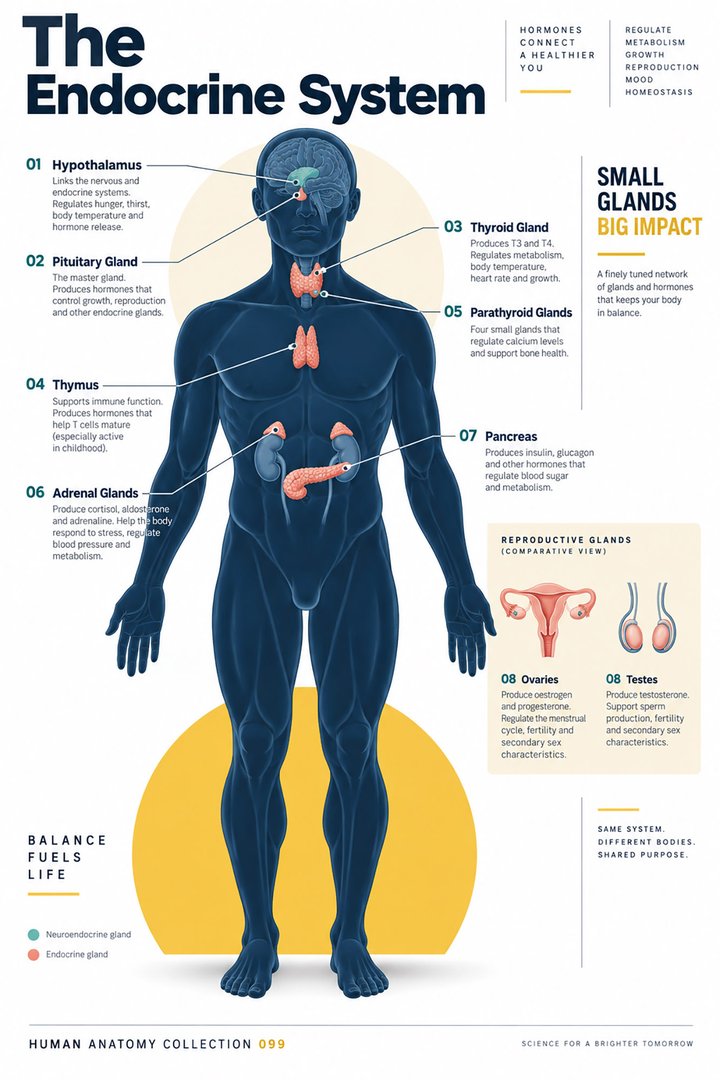 Human Endocrine System ↗
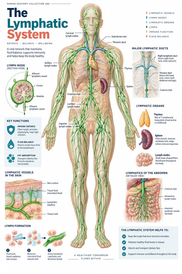 Human Lymphatic System ↗
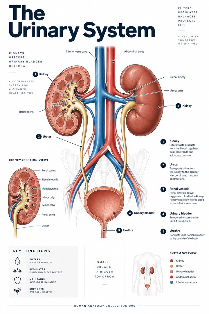 Human Urinary System ↗
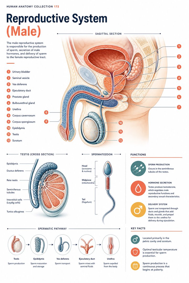 Male Reproductive System ↗
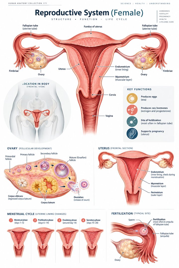 Female Reproductive System ↗
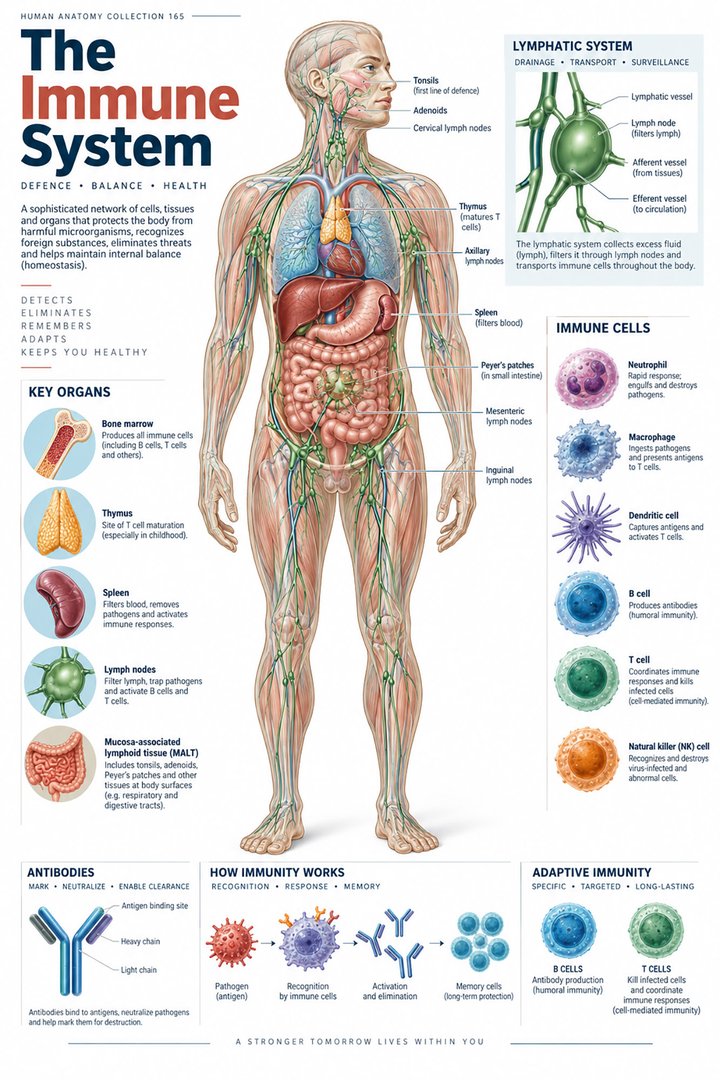 Human Immune System ↗Central Nervous System ↗
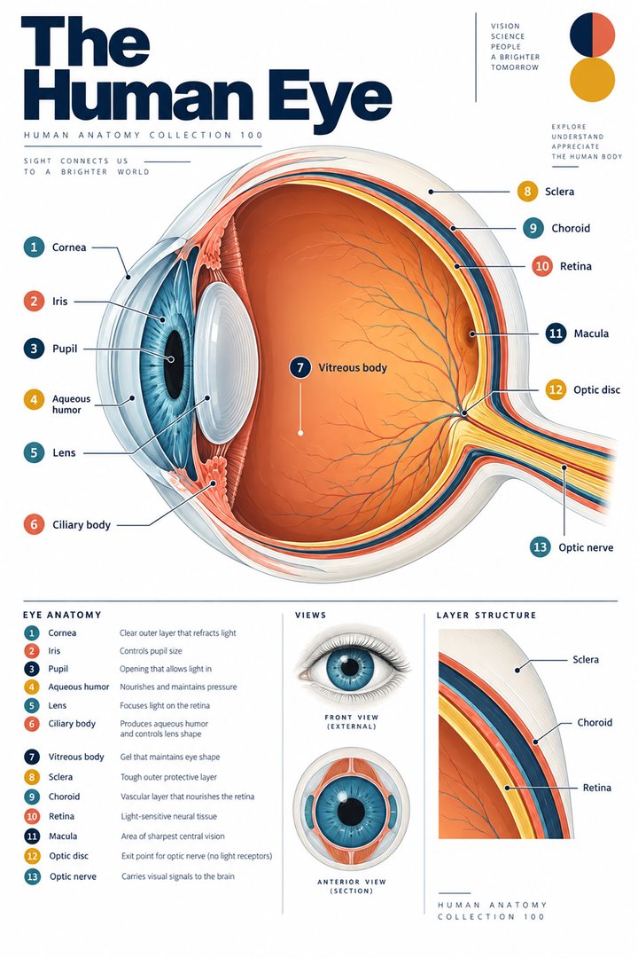 Human Eye Structure ↗
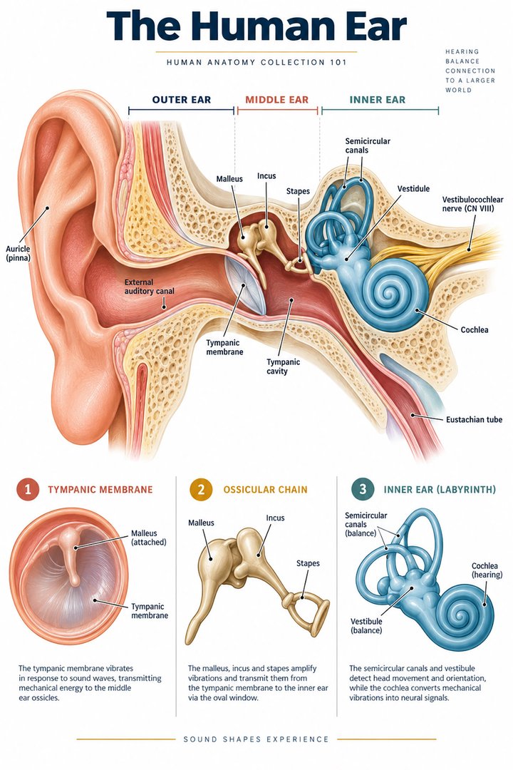 Human Ear Structure ↗
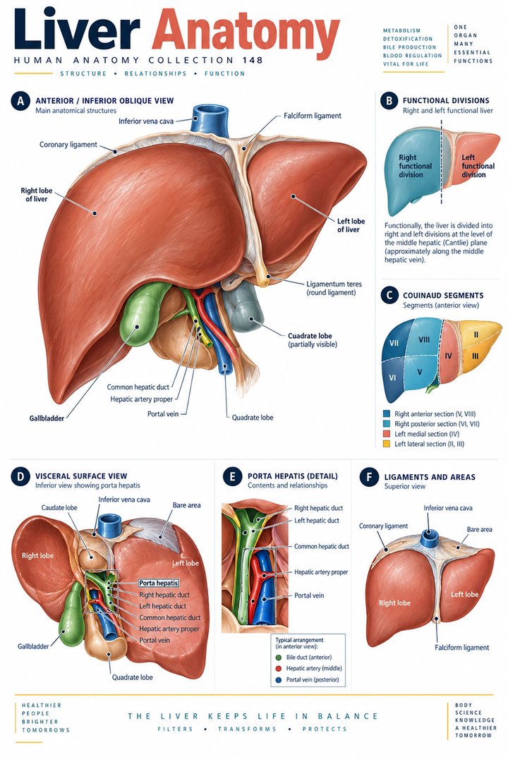 Human Liver Function ↗
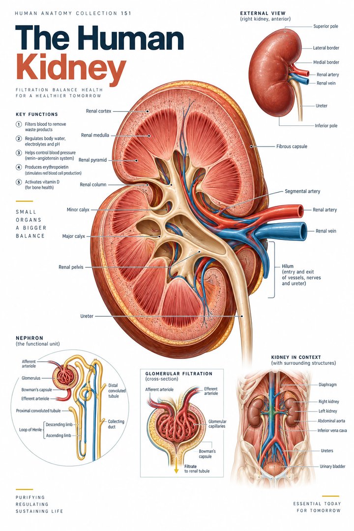 Human Kidney Anatomy ↗
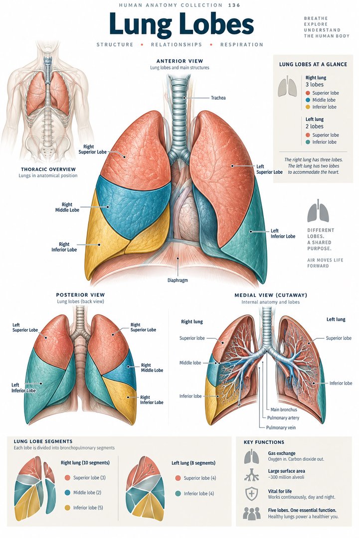 Human Lung Anatomy ↗
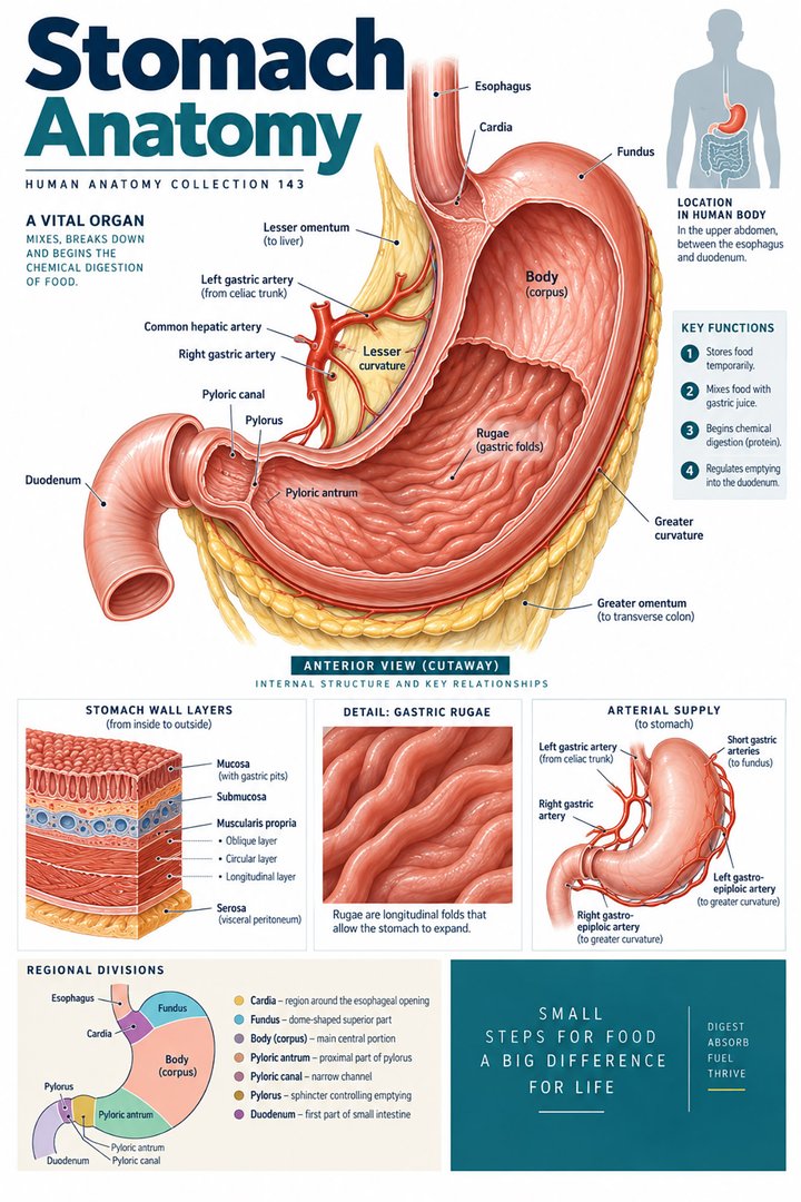 Human Stomach Structure ↗
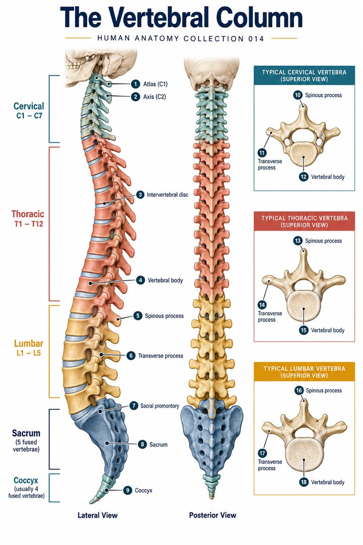 Human Spine Anatomy ↗
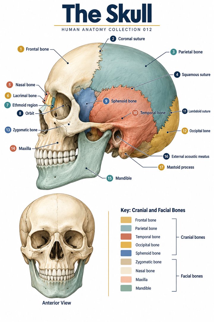 Human Skull Anatomy ↗
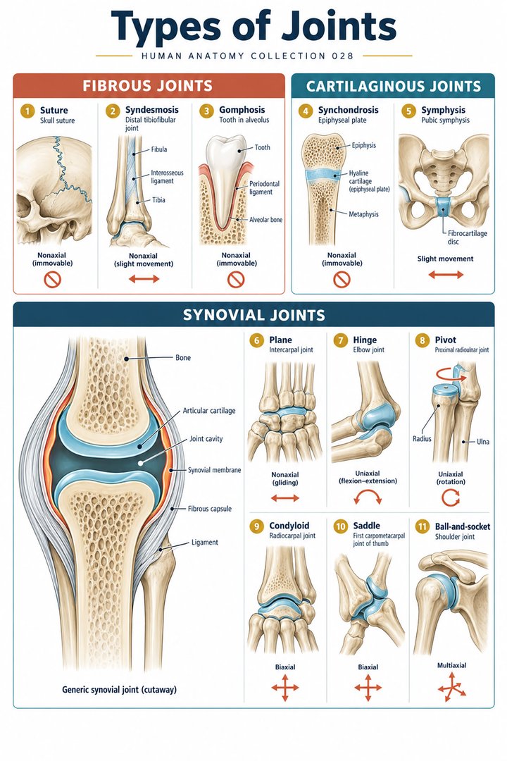 Human Joint Types ↗
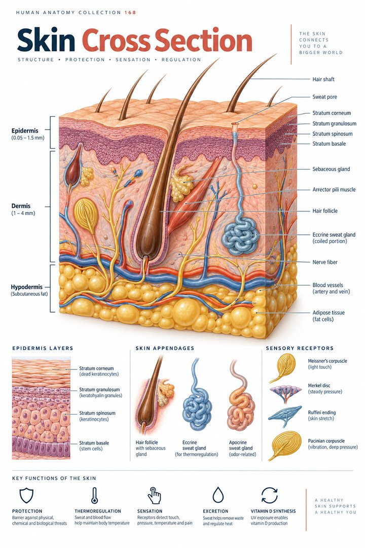 Human Skin Layers ↗
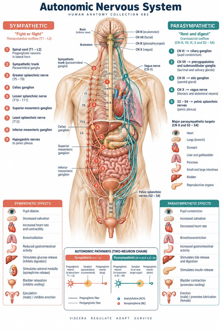 Autonomic Nervous System ↗
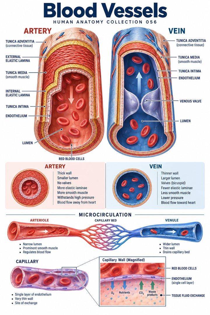 Human Blood Vessels ↗
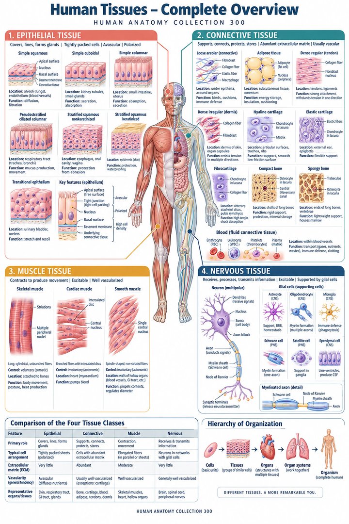 Human Body Tissues ↗
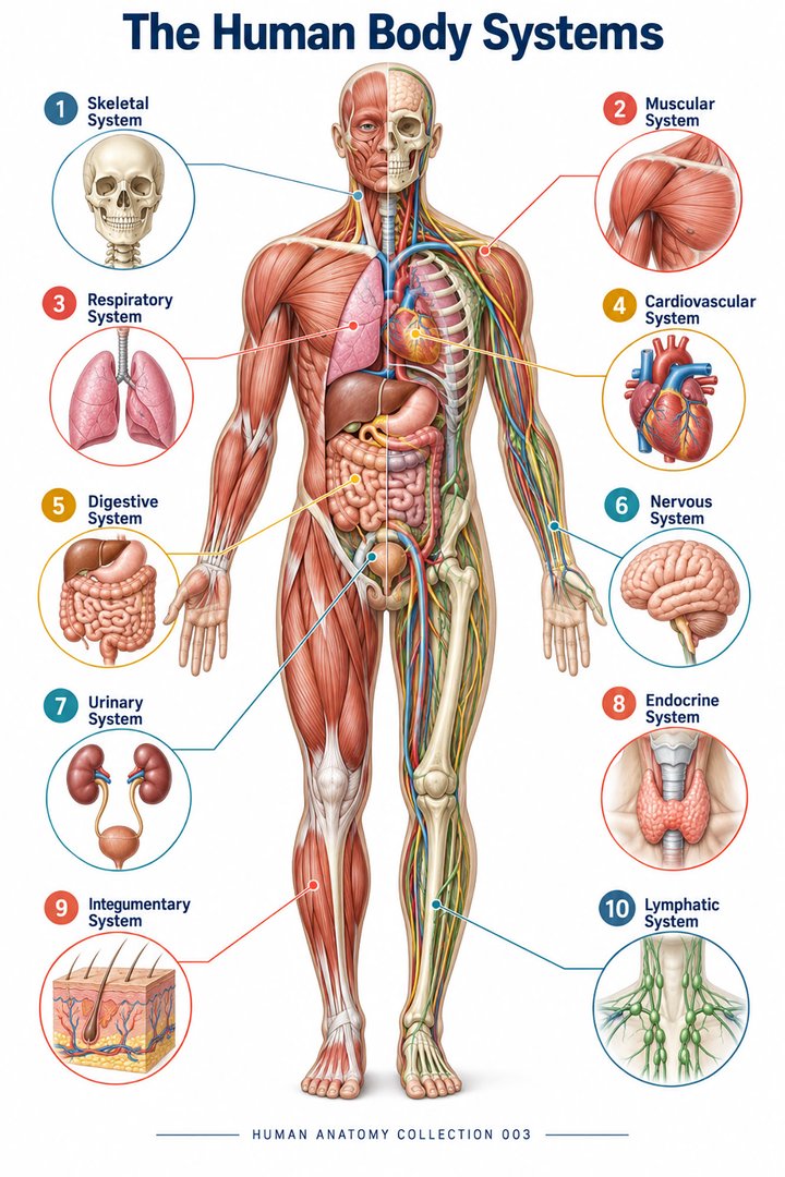 Human Body Systems ↗
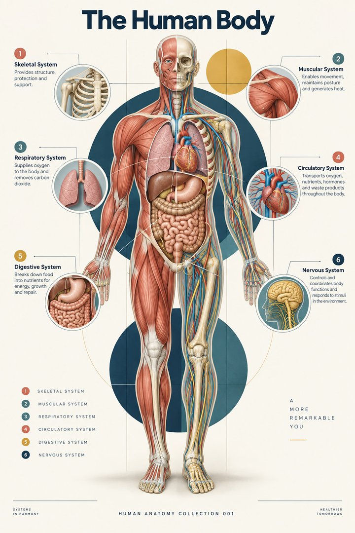 Human Body Facts ↗Human Anatomy and Physiology ↗
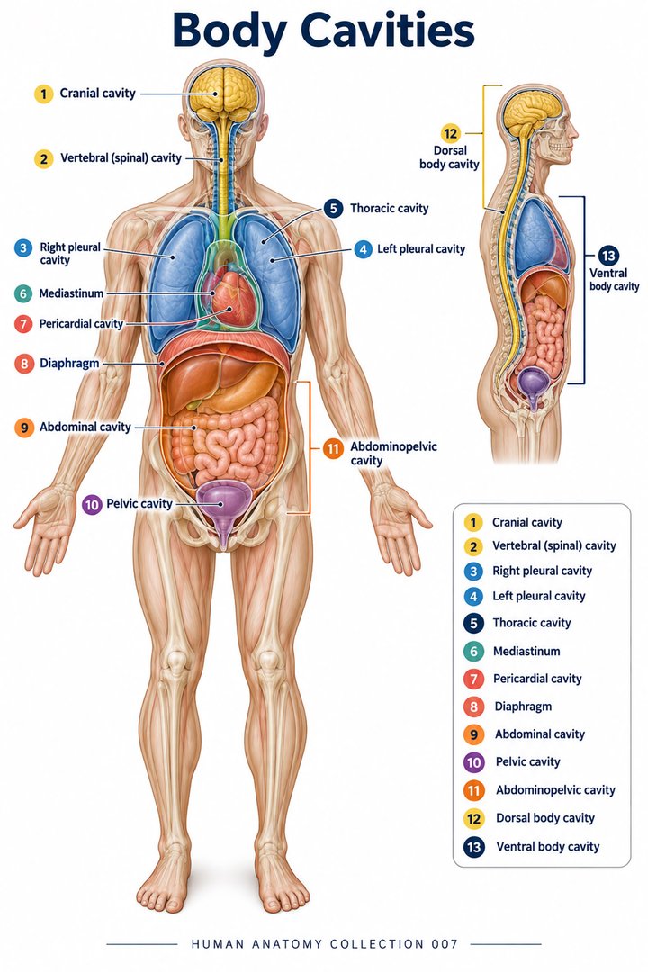 Human Body Cavities ↗Human Torso Anatomy ↗Human Digestive Tract ↗
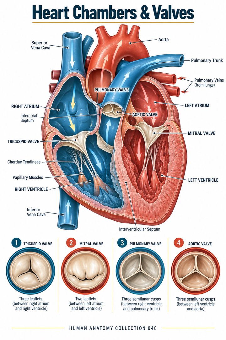 Human Heart Chambers ↗Human Organ Placement ↗
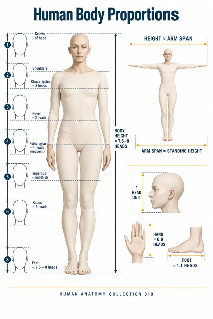 Human Body Proportions ↗
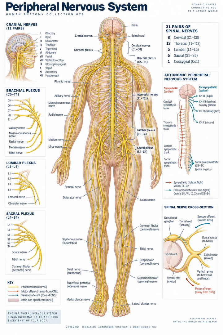 Peripheral Nervous System ↗
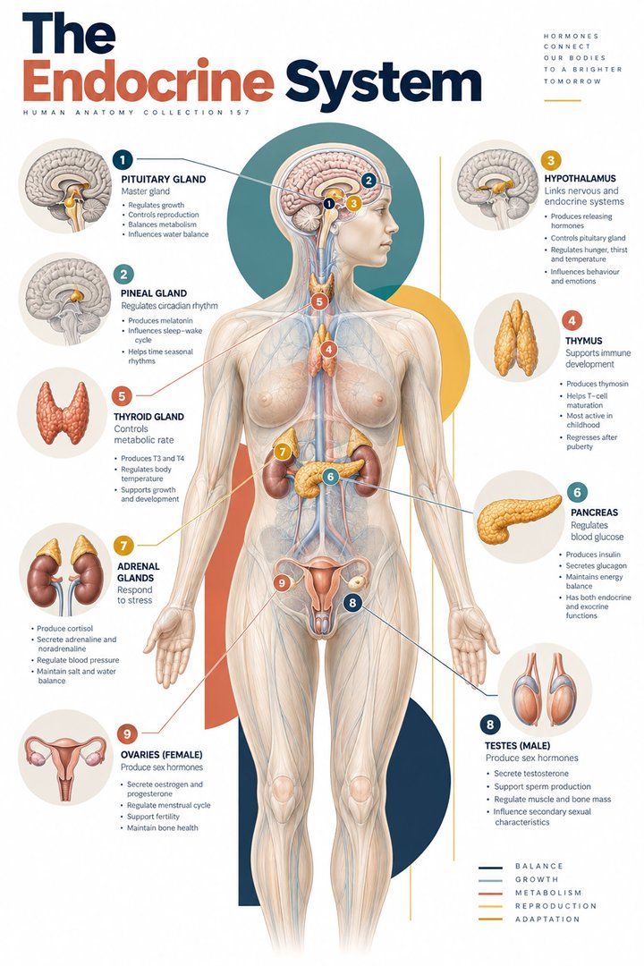 Human Endocrine Glands ↗
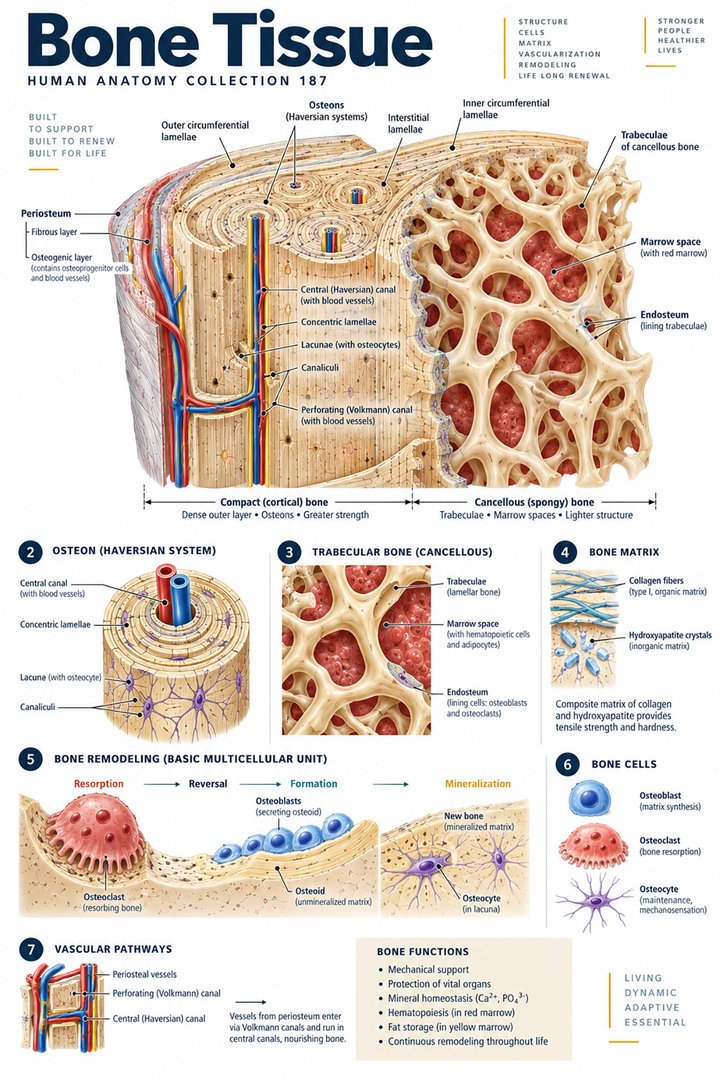 Human Bone Density ↗Human Blood Circulation ↗
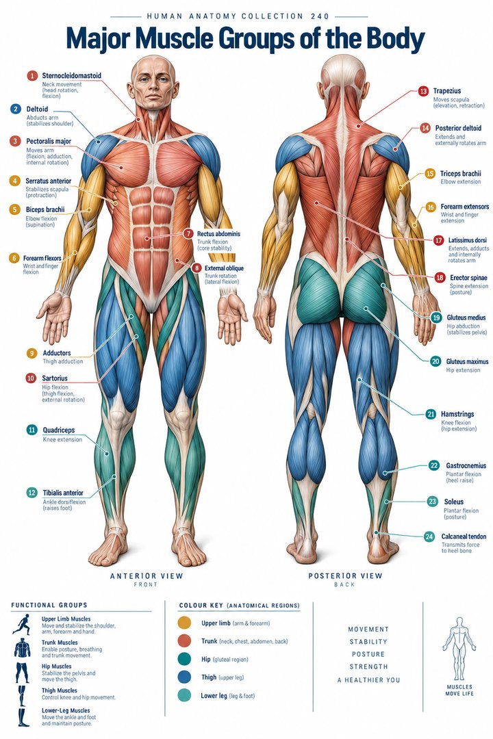 Human Muscle Groups ↗
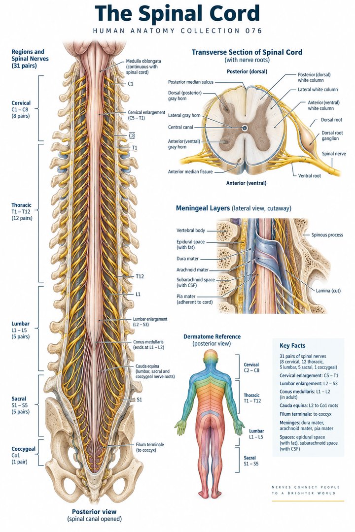 Human Spinal Cord ↗
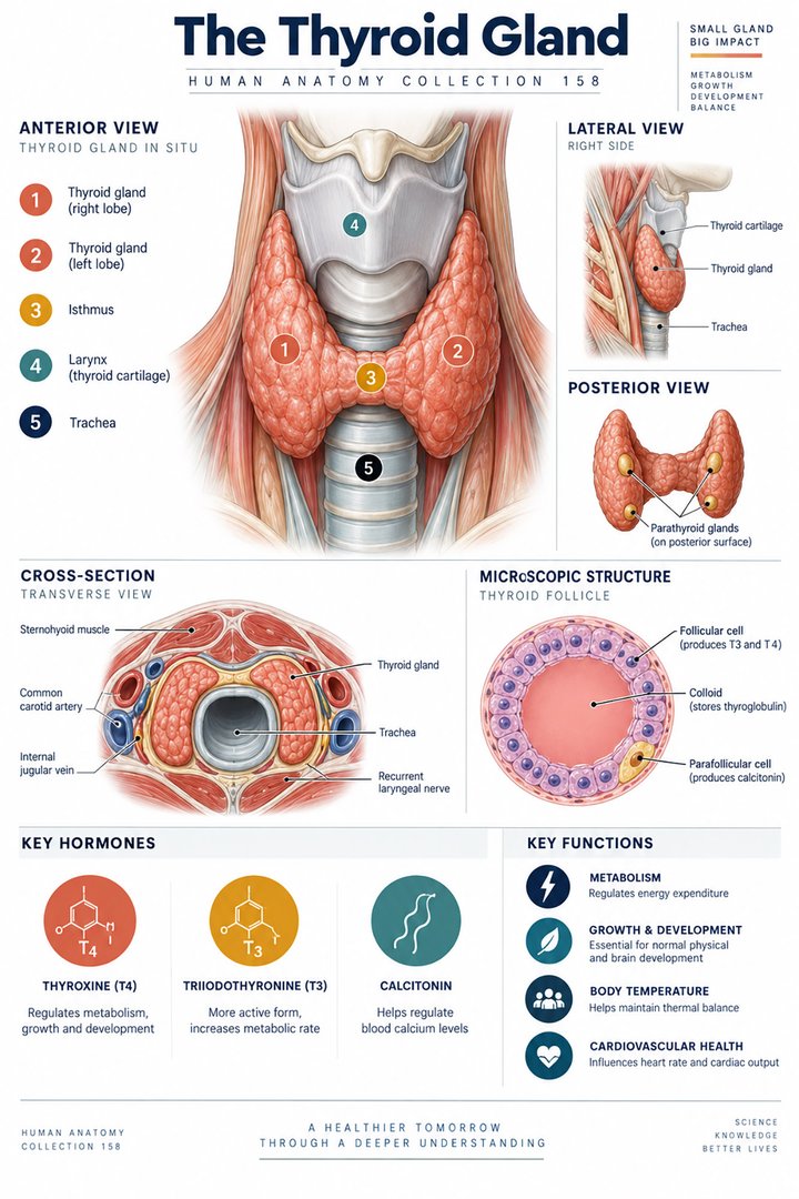 Human Thyroid Gland ↗
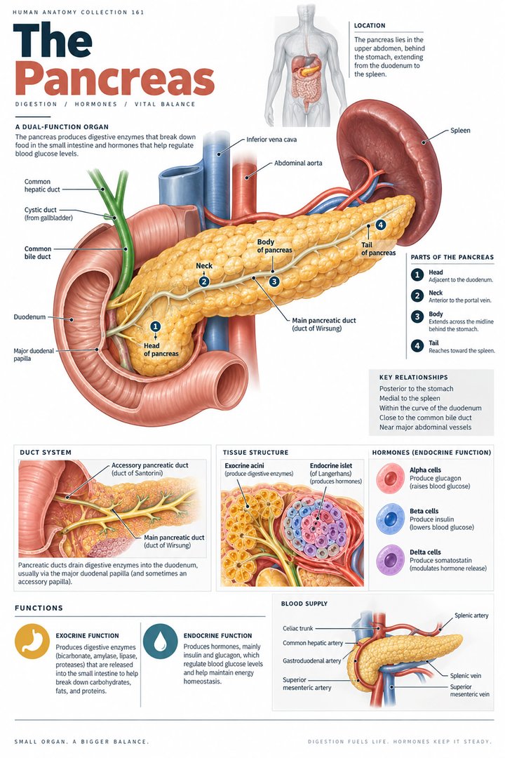 Human Pancreas Function ↗
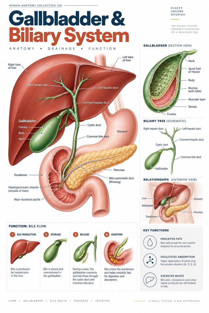 Human Gallbladder Anatomy ↗