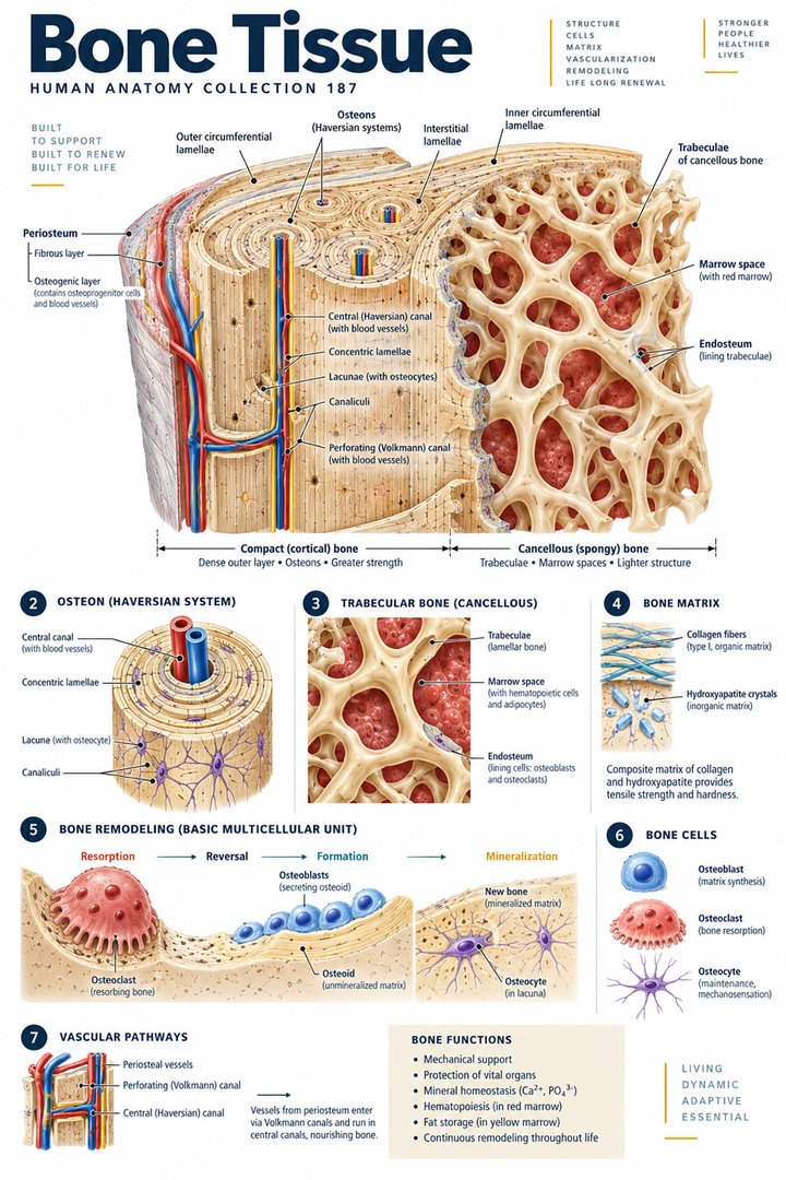
Bone Tissue
Individual poster · 300 DPI print files
Regular $9.90
Explore poster ↗$4.90USD
Offer price · Save $5.00Individual poster · 300 DPI print files
Reduced web preview. Separate high-resolution print files are supplied with the digital download.
ANATOMY 187
A modern anatomy illustration exploring bone tissue, designed for educational and decorative use.
Purchases are not available yet. Checkout and download delivery are being prepared.
Sold by Kunstolymp.de · Germany
Compare prices & the complete collection ↗This is a DIGITAL DOWNLOAD. No physical product will be shipped. After purchase, download your files and print them at home, at a local print shop, or through an online professional printing service.
The separate 300 DPI print files are ideal for 24×36-inch posters (approximately 60×90 cm). ISO and 5×7 formats are included. The white border lets you adjust the layout or trim to fit other frame sizes; check the preview before printing to keep the illustration and labels visible.
The planned bundle delivery includes a PDF information sheet with a link to the relevant Google Drive folder and download instructions. The store is currently in preview and purchases are not yet available.
A science-inspired choice for a student’s study corner, an educator’s classroom, or an anatomy enthusiast’s wall. Final licensing terms will apply.
CLINICAL CONTEXT
Bone is living connective tissue rather than an inert scaffold. Its collagen-rich matrix provides flexibility, while mineral deposition contributes hardness. Compact cortical bone forms a dense outer shell; trabecular bone creates an internal network that distributes load. Blood vessels supply bone cells, and marrow spaces support blood-cell production. Osteoblasts form new matrix, osteoclasts resorb bone, and osteocytes help coordinate responses to mechanical loading. Continual remodelling replaces old tissue and supports repair. Bone strength therefore depends on tissue quality, architecture, and mineral content together. A tissue diagram helps explain these relationships, but cannot establish a patient's bone density or fracture risk.
Osteoporosis weakens bone and increases fracture risk, often without symptoms until a fracture occurs. Vertebral fractures may cause back pain, height loss, or altered posture; hip and wrist fractures are also important concerns. Osteomalacia involves inadequate mineralisation of adult bone and can cause diffuse pain or muscle weakness, often in association with vitamin D deficiency. Paget's disease produces abnormal remodelling in affected bones, which may become enlarged, deformed, or painful. Osteomyelitis is bone infection and may cause focal pain, swelling, warmth, and fever. Bone pain is not a diagnosis: injury, infection, metabolic disease, and other causes require different assessment. Ask about falls, previous fractures, nutrition, mobility, long-term glucocorticoids, and relevant medical history.
Assessment combines symptoms, examination, prior fractures, medicines, and fracture-risk factors. Dual-energy X-ray absorptiometry (DXA), usually at the hip and spine, measures bone mineral density and helps guide osteoporosis assessment. Suspected fracture or focal disease may need other imaging. Blood tests are selected to investigate underlying causes rather than diagnose all bone problems from one result. Suspected infection requires prompt evaluation and may involve blood tests, imaging, and sampling. Persistent localised pain should be investigated rather than attributed automatically to ageing.
Osteoporosis treatment combines fracture prevention with selected medicines that reduce bone resorption or promote formation. Choice and follow-up depend on risk and individual circumstances. Osteomalacia treatment corrects the cause and appropriate nutrient deficiencies; Paget's disease may need specialist treatment for symptoms or complications. Osteomyelitis usually requires antibiotics and may require surgery. Fracture care and rehabilitation aim to restore function and reduce further injury. Adequate nutrition, safe strength and weight-bearing activity, and fall prevention support treatment; exercise must be adapted when fractures or severe fragility are present.
Physiotherapy-guided movement, balance work, and practical home changes can support independence. Relaxation may help coping with pain but does not correct infection or abnormal mineralisation. Calcium and vitamin D intake should be reviewed according to individual needs; supplements are appropriate when indicated rather than taken automatically at high doses. Discuss products and interactions with a clinician. Herbal remedies and detox regimens should not replace prescribed bone treatment. Avoid forceful manipulation of a painful area or suspected fracture. After a low-trauma fracture, arrange bone-health assessment as well as recovery care.
Severe pain, deformity, or inability to use a limb after injury needs urgent assessment. Localised bone pain with fever, redness, or swelling requires prompt review for possible infection. An open injury with exposed bone or signs of serious systemic illness requires emergency care. Persistent or worsening focal pain and unexplained height loss should also be assessed. Explain safe activity, the follow-up plan, and reasons to seek help. This guide supports clinical learning and does not replace an individual examination.
Further reading: NIAMS: bone health · NIAMS: osteoporosis · NIAMS: diagnosis and treatment · NIAMS: Paget's disease · NHS: osteomalacia · NHS: osteomyelitis
Explain the working diagnosis, important alternatives, planned tests, expected course, and specific safety-netting in language the patient can use. Invite questions about treatment goals, capacity, cost, culture, and complementary therapies. Use a professional interpreter when language access could affect consent or understanding, and confirm clear arrangements for timely clinical review in writing. Document the reasoning, reassess response to treatment, and coordinate care across professions when illness affects more than one system. Teaching anatomy is most useful when it supports this partnership: accurate localisation, respectful listening, timely escalation, and a plan that remains responsive to the individual patient.
HUMAN ANATOMY COLLECTION · 001—300
A closer look at what makes us human. Discover anatomy posters for your study space, classroom, and everyday curiosity.
DIGITAL DOWNLOADS · 300 DPI PRINT FILES
EXPLORE BY SYSTEM
LEARN & EXPLORE
Detailed English guides with Latin terminology, diagrams, real images, and clinical learning connections.
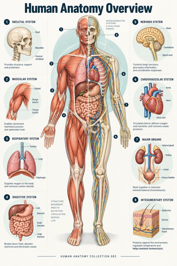 Human Anatomy Diagrams ↗
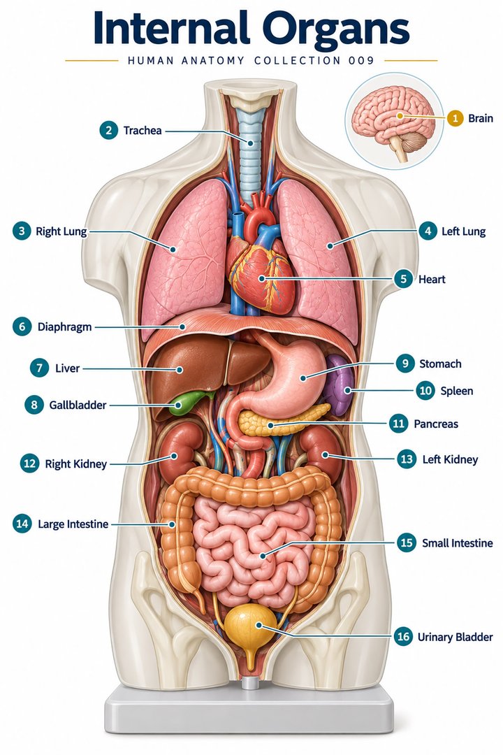 Human Body Organs ↗
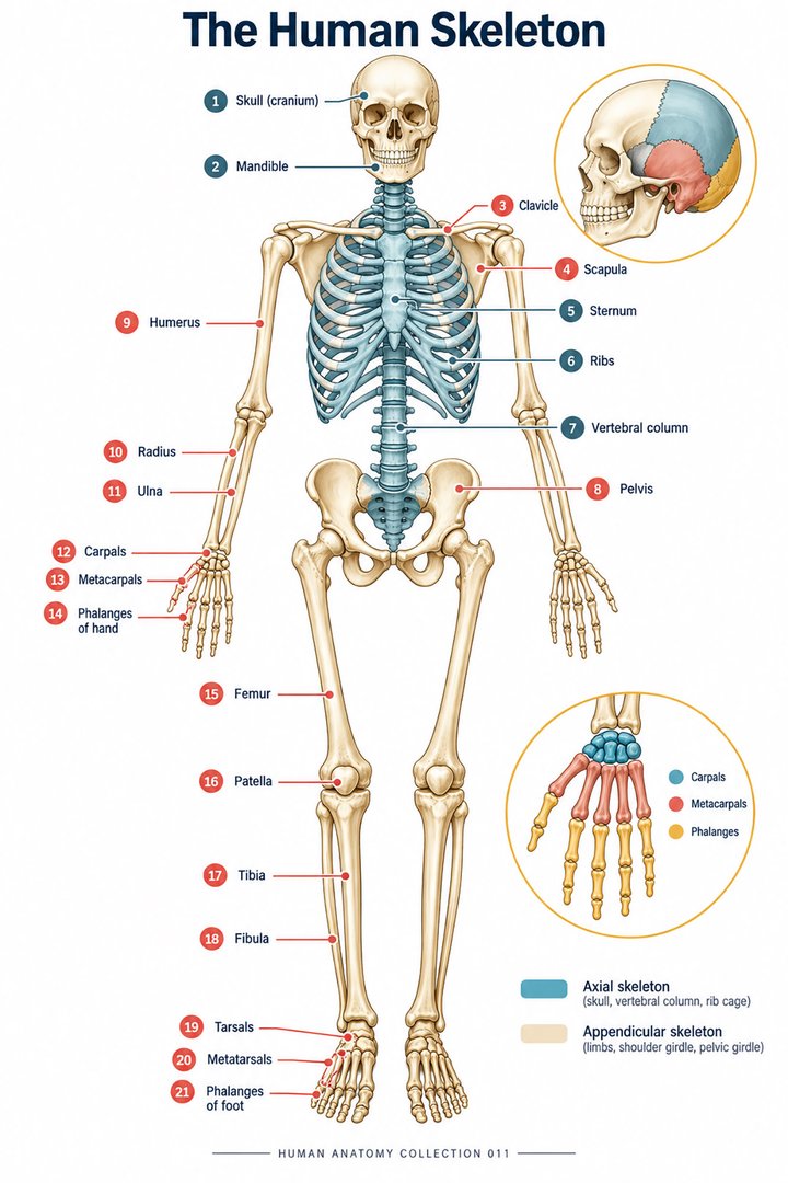 Skeletal System Anatomy ↗
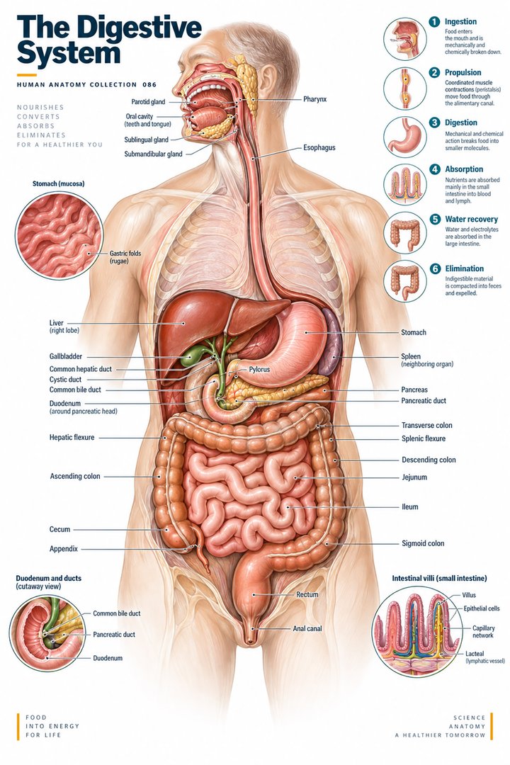 Human Digestive System ↗
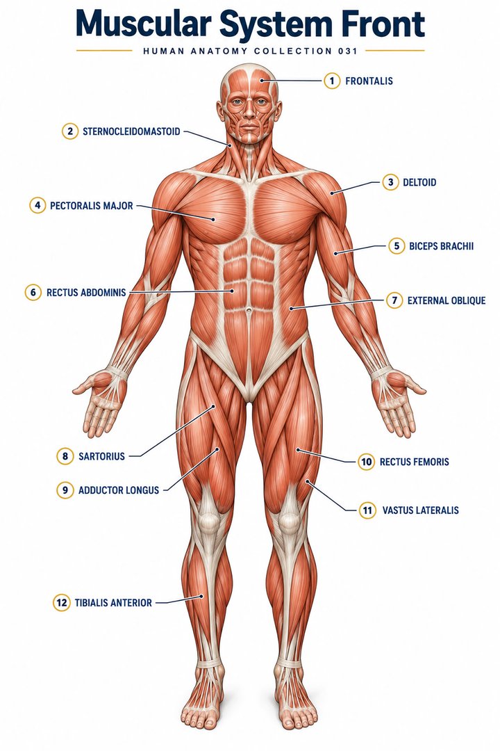 Muscular System Overview ↗
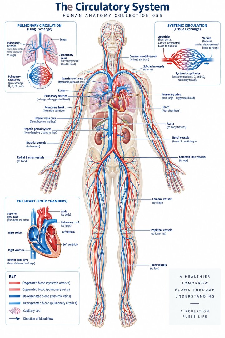 Human Circulatory System ↗
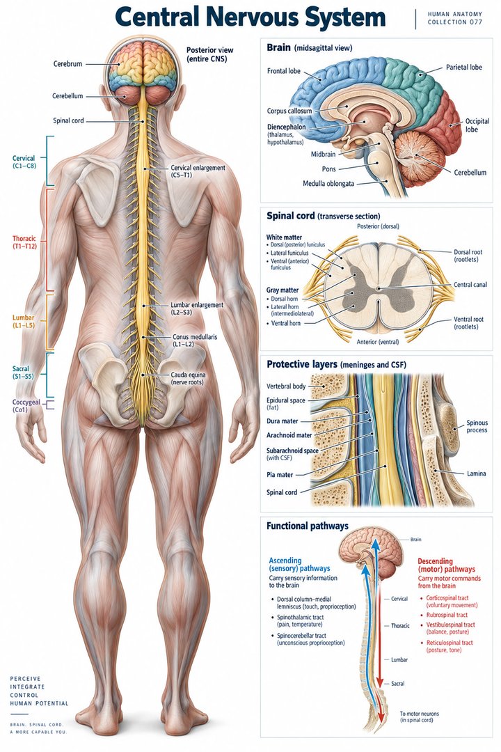 Human Nervous System ↗
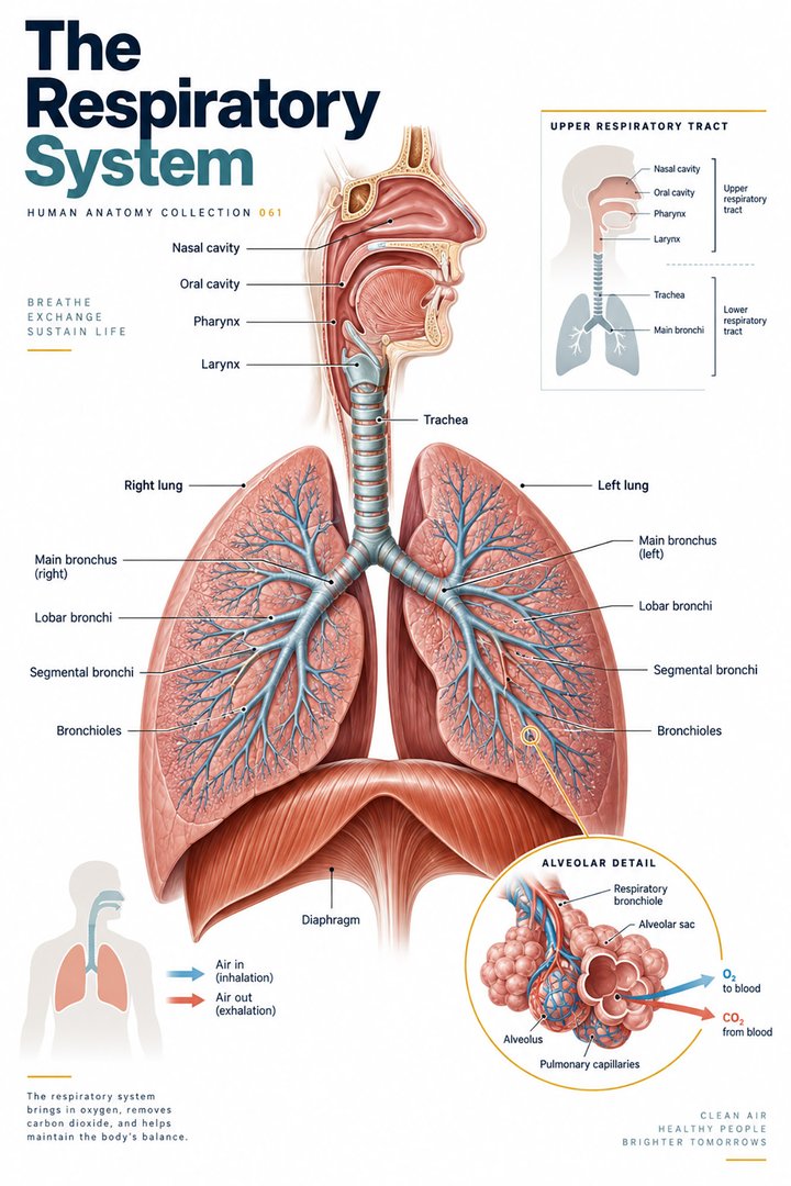 Respiratory System Functions ↗
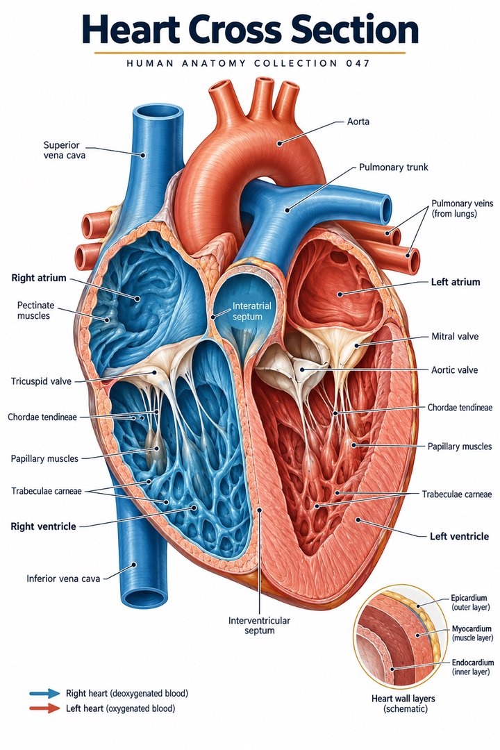 Human Heart Structure ↗
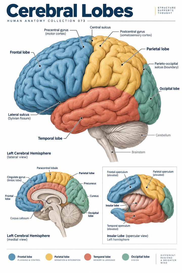 Human Brain Lobes ↗
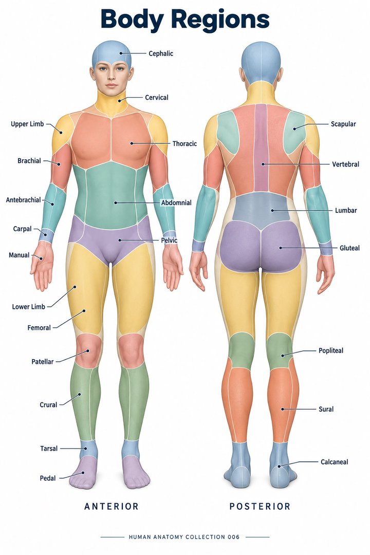 Human Body Parts ↗
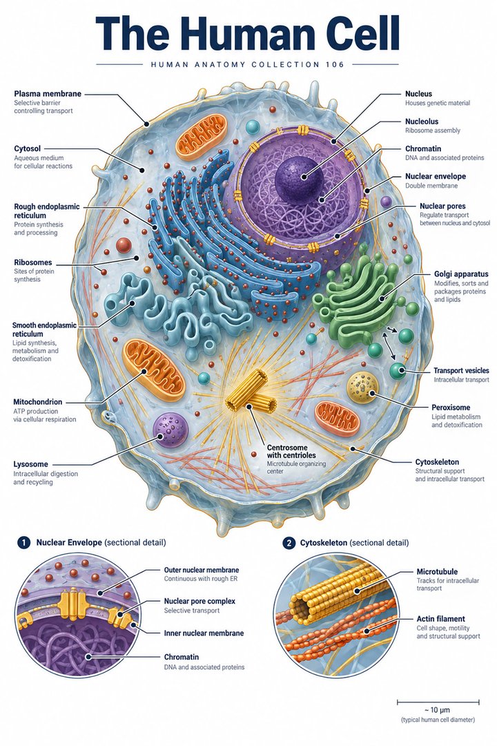 Human Cell Structure ↗
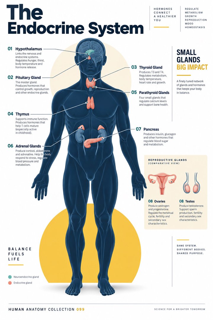 Human Endocrine System ↗
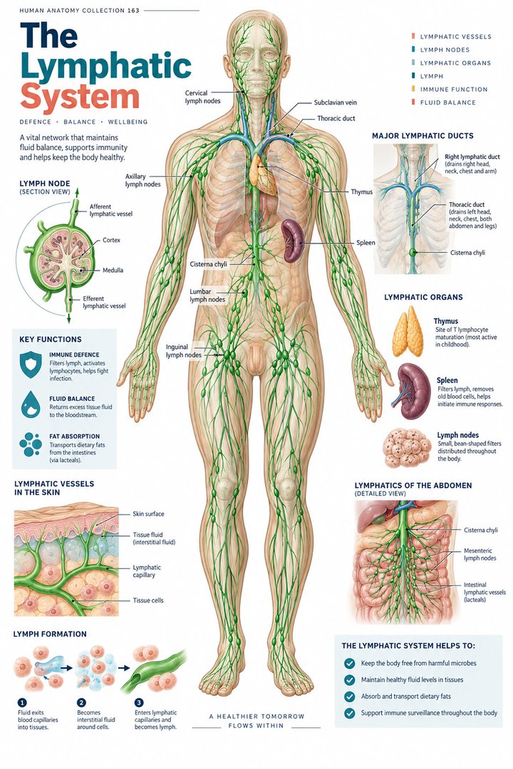 Human Lymphatic System ↗
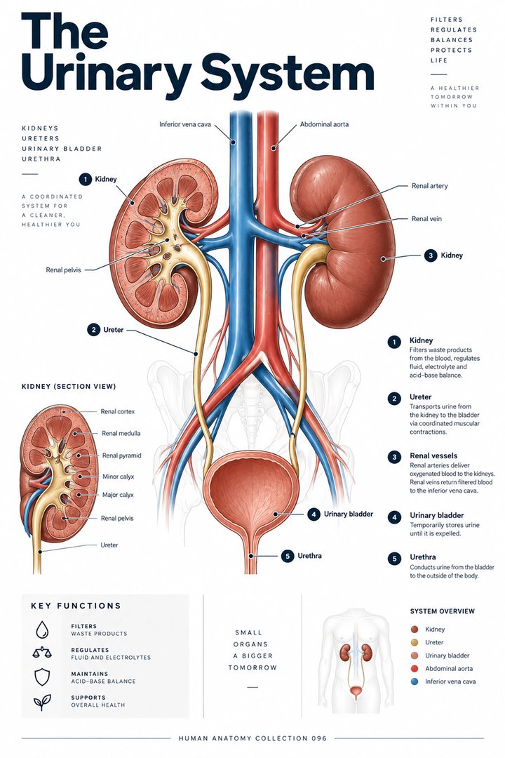 Human Urinary System ↗
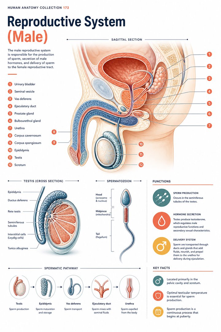 Male Reproductive System ↗
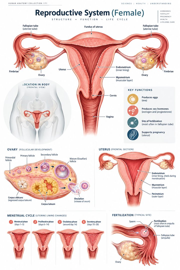 Female Reproductive System ↗
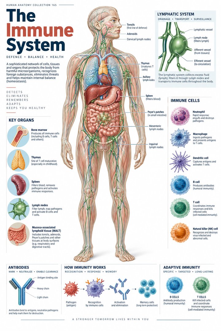 Human Immune System ↗Central Nervous System ↗
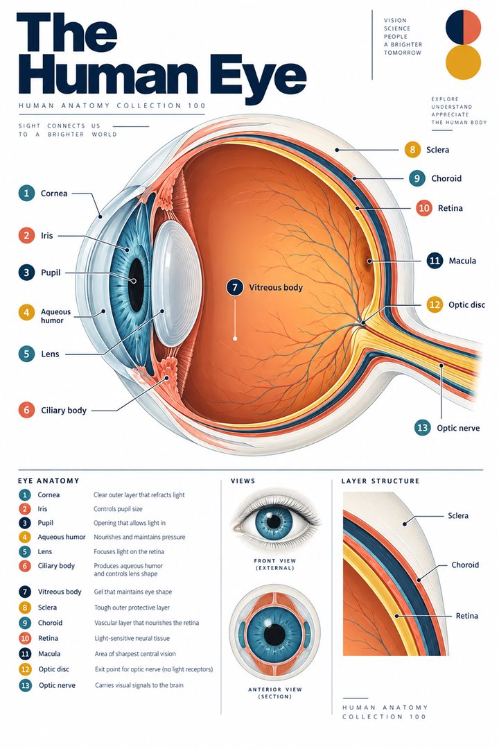 Human Eye Structure ↗
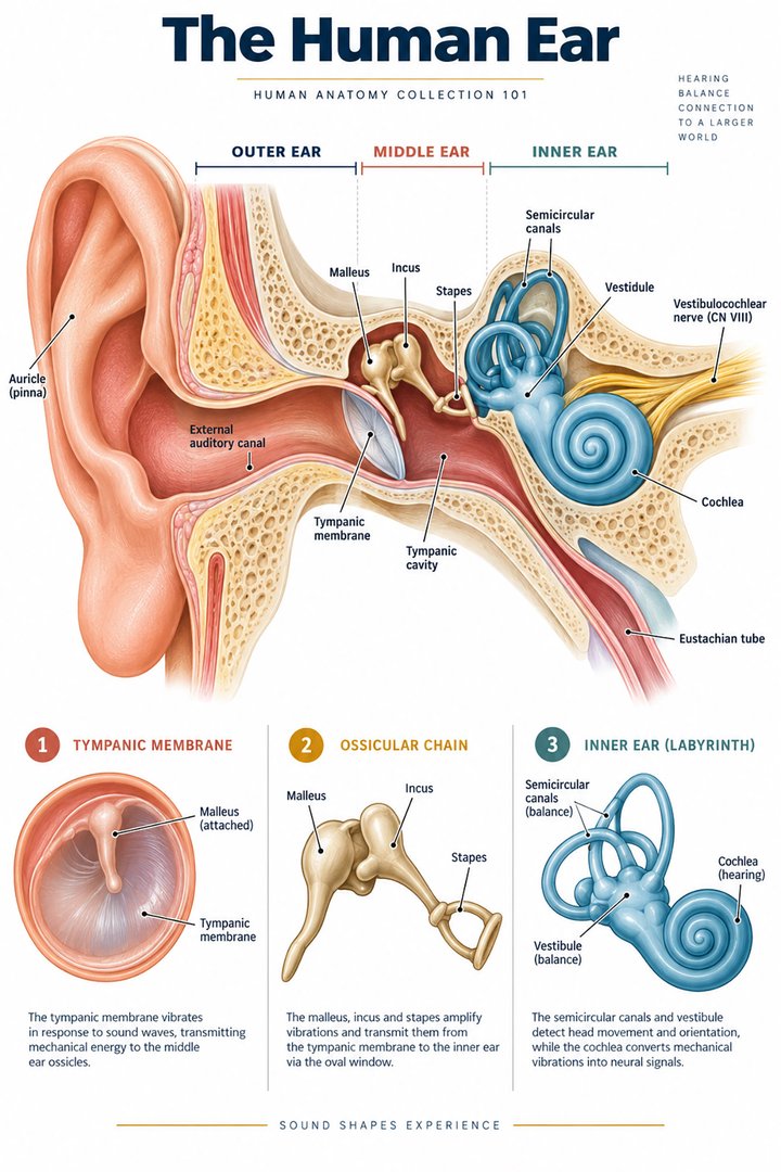 Human Ear Structure ↗
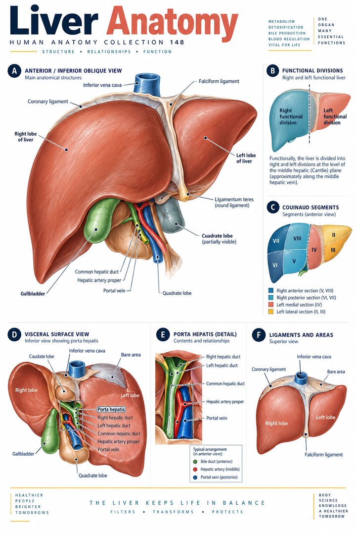 Human Liver Function ↗
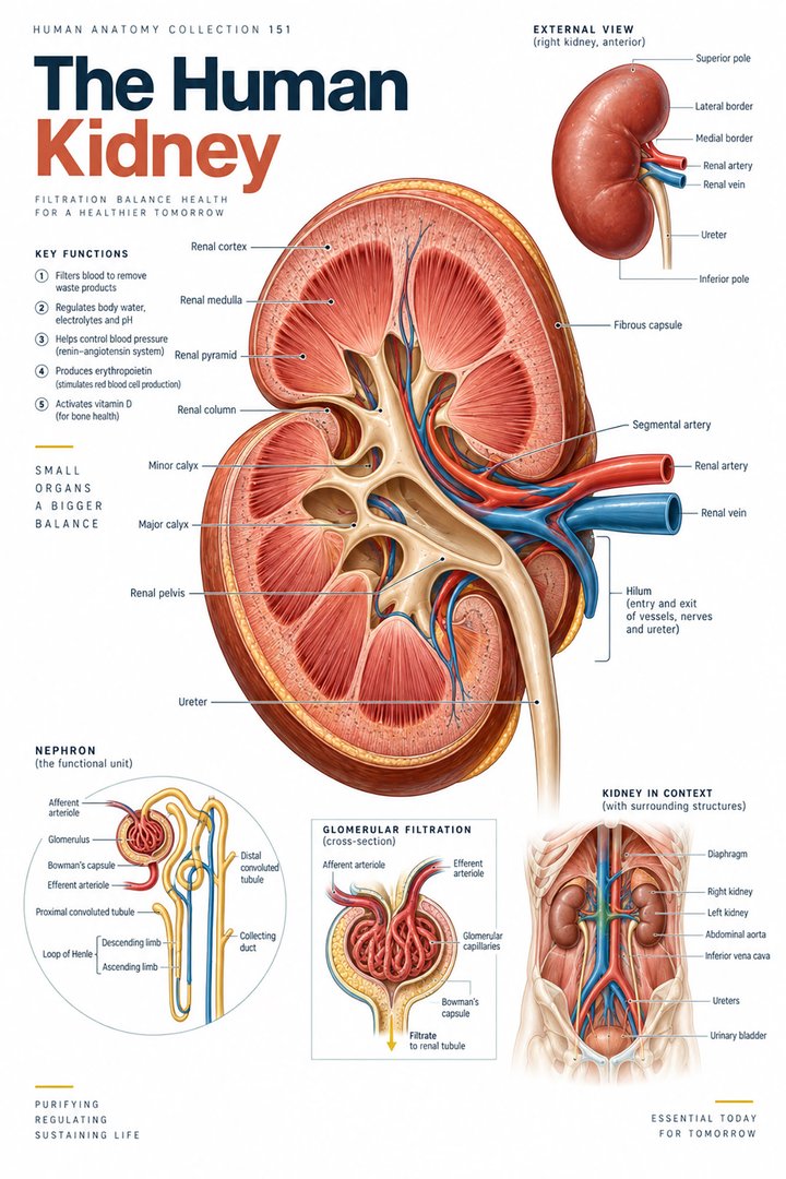 Human Kidney Anatomy ↗
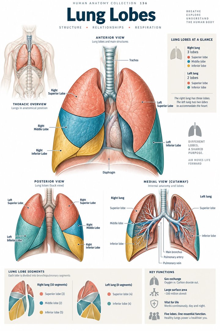 Human Lung Anatomy ↗
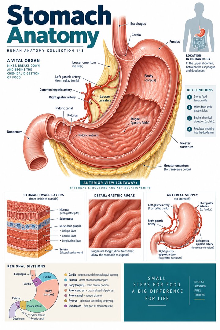 Human Stomach Structure ↗
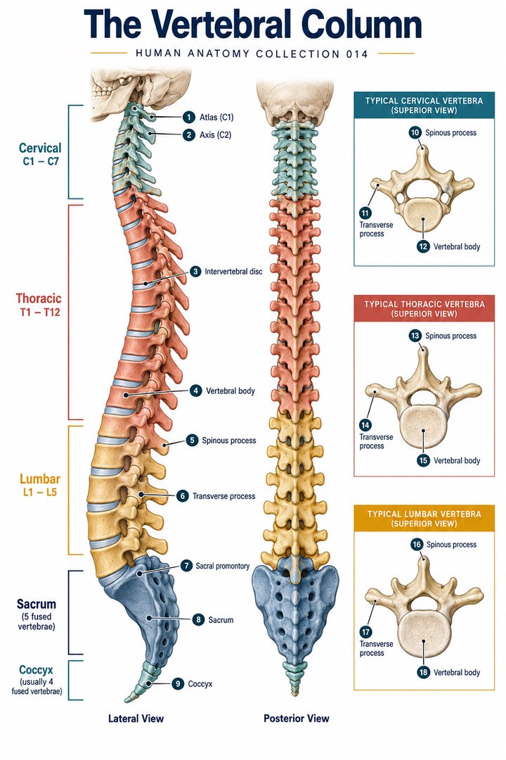 Human Spine Anatomy ↗
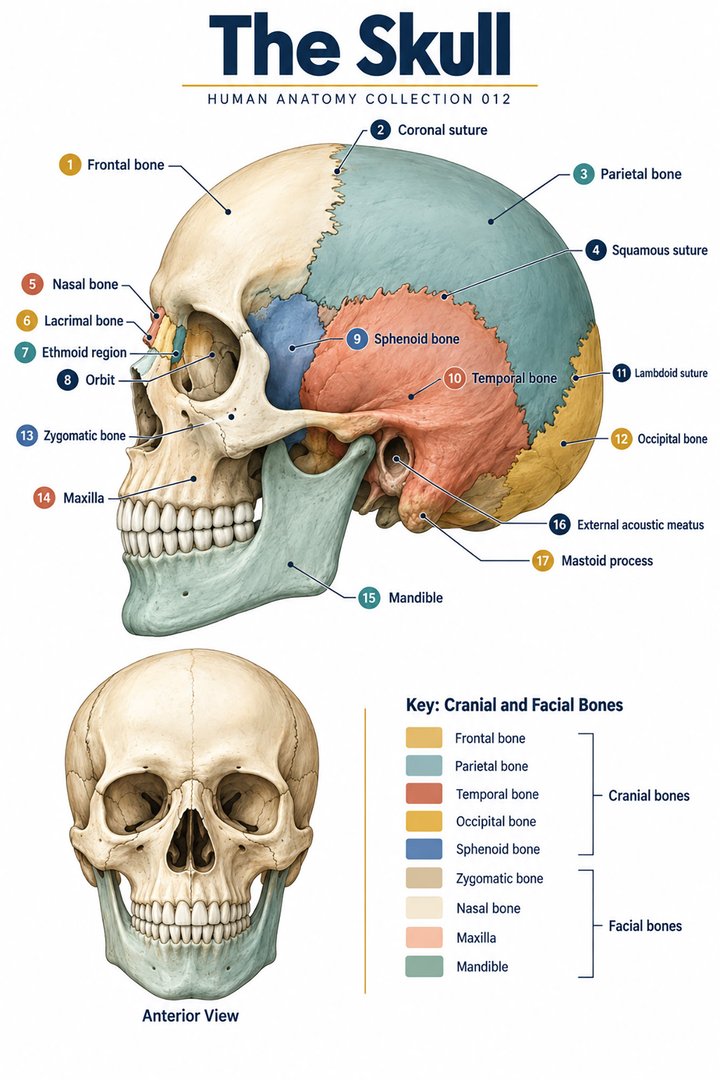 Human Skull Anatomy ↗
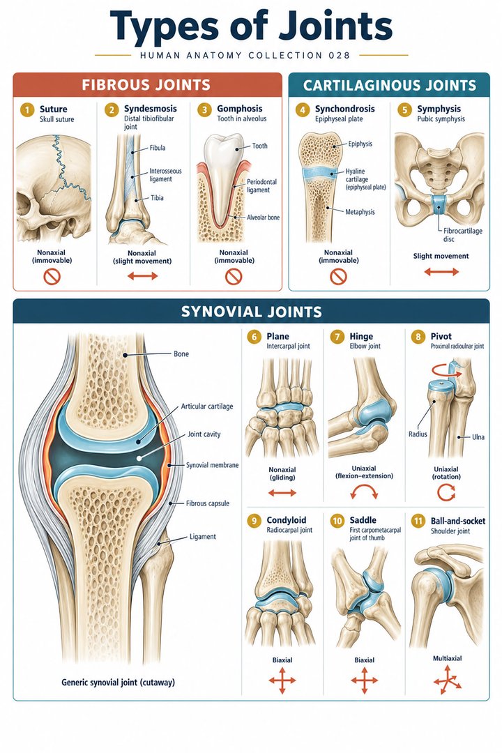 Human Joint Types ↗
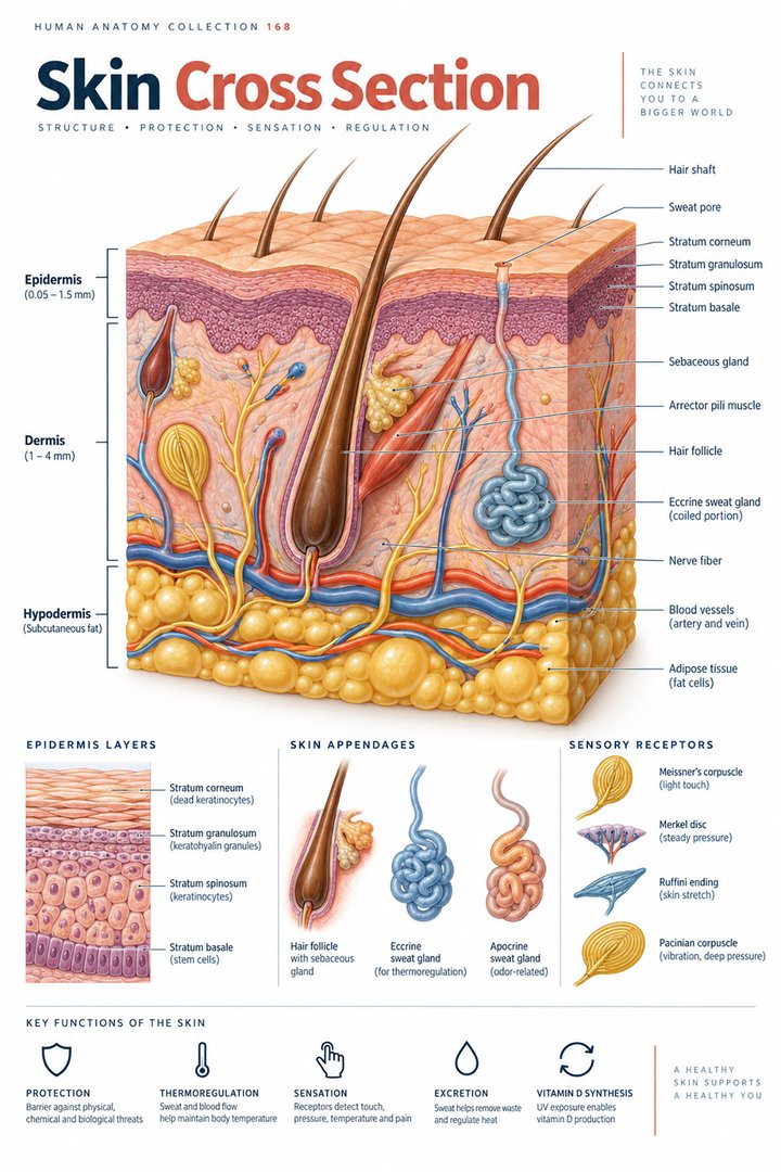 Human Skin Layers ↗
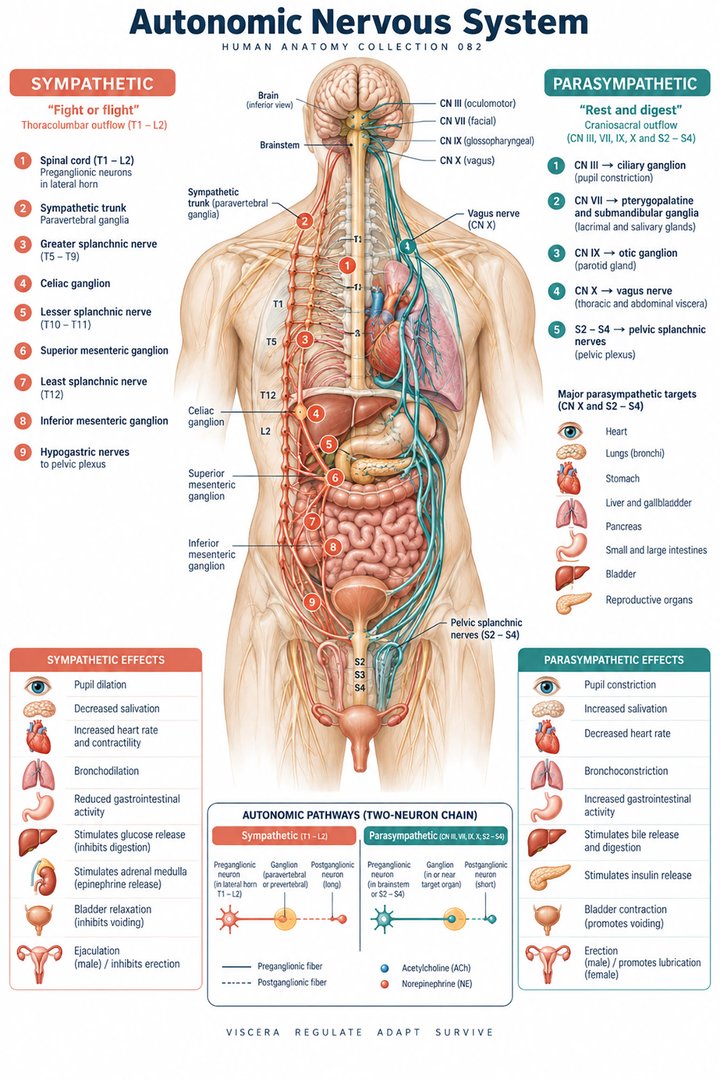 Autonomic Nervous System ↗
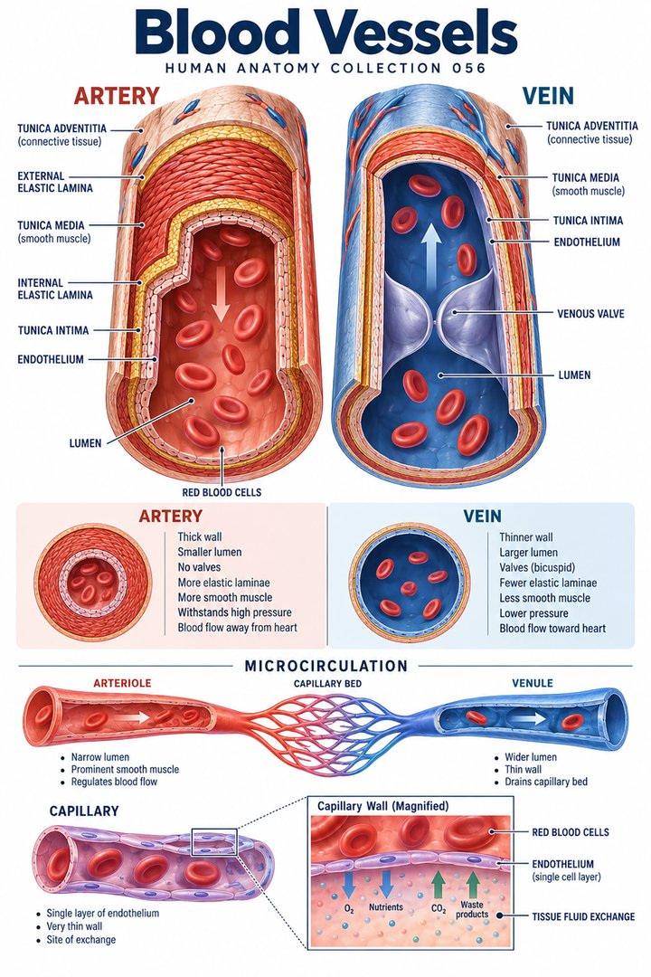 Human Blood Vessels ↗
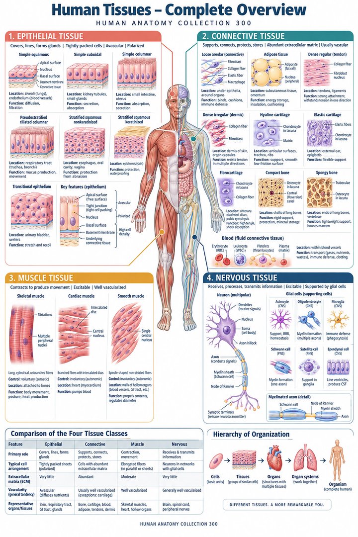 Human Body Tissues ↗
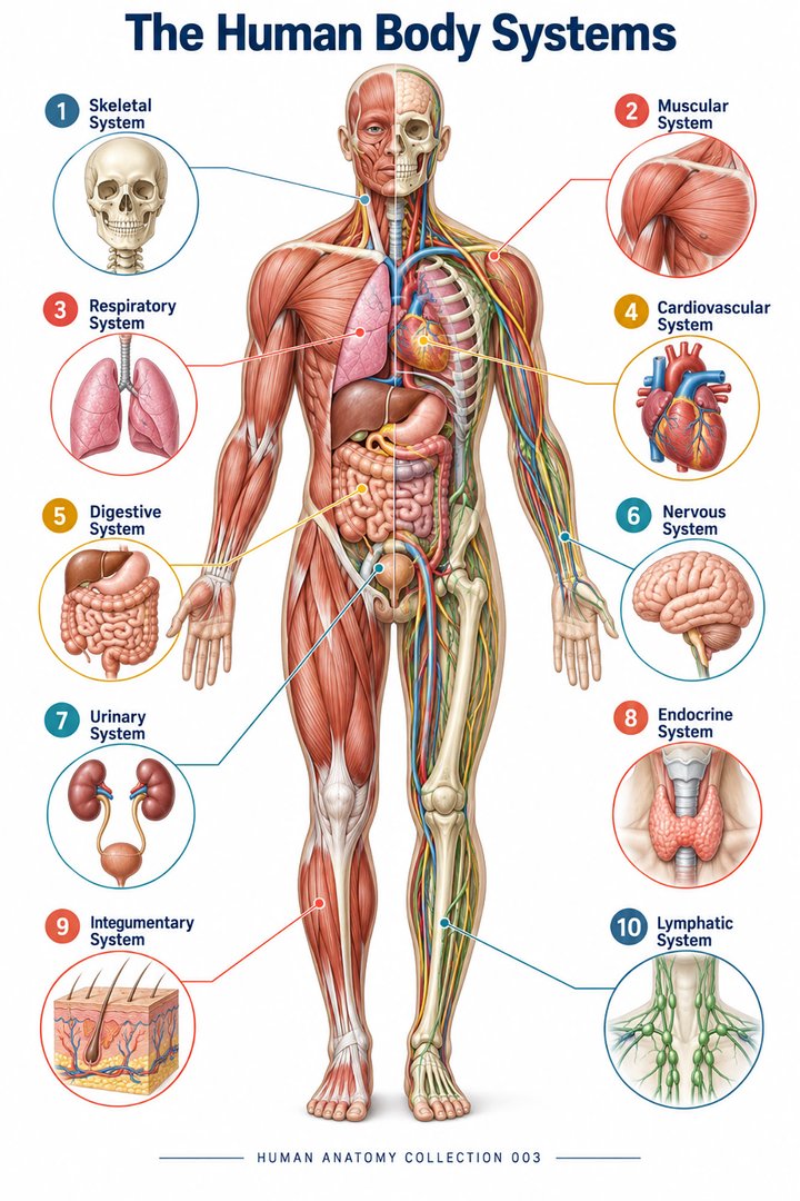 Human Body Systems ↗
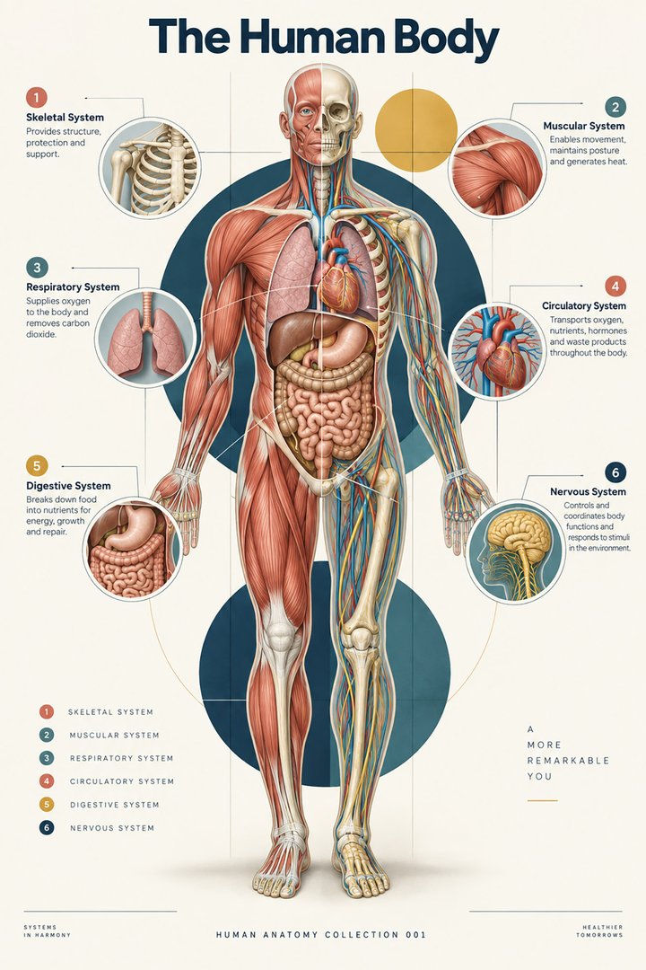 Human Body Facts ↗Human Anatomy and Physiology ↗
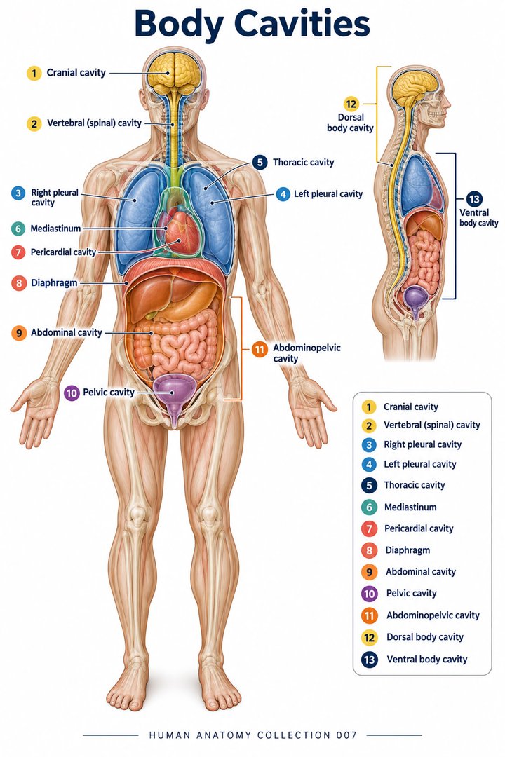 Human Body Cavities ↗Human Torso Anatomy ↗Human Digestive Tract ↗
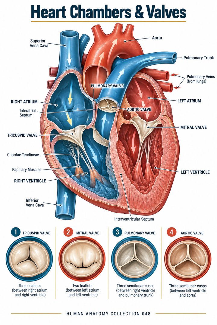 Human Heart Chambers ↗Human Organ Placement ↗
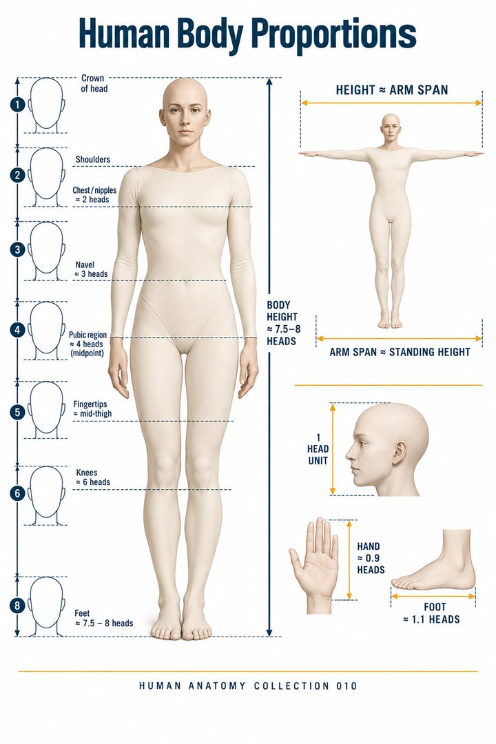 Human Body Proportions ↗
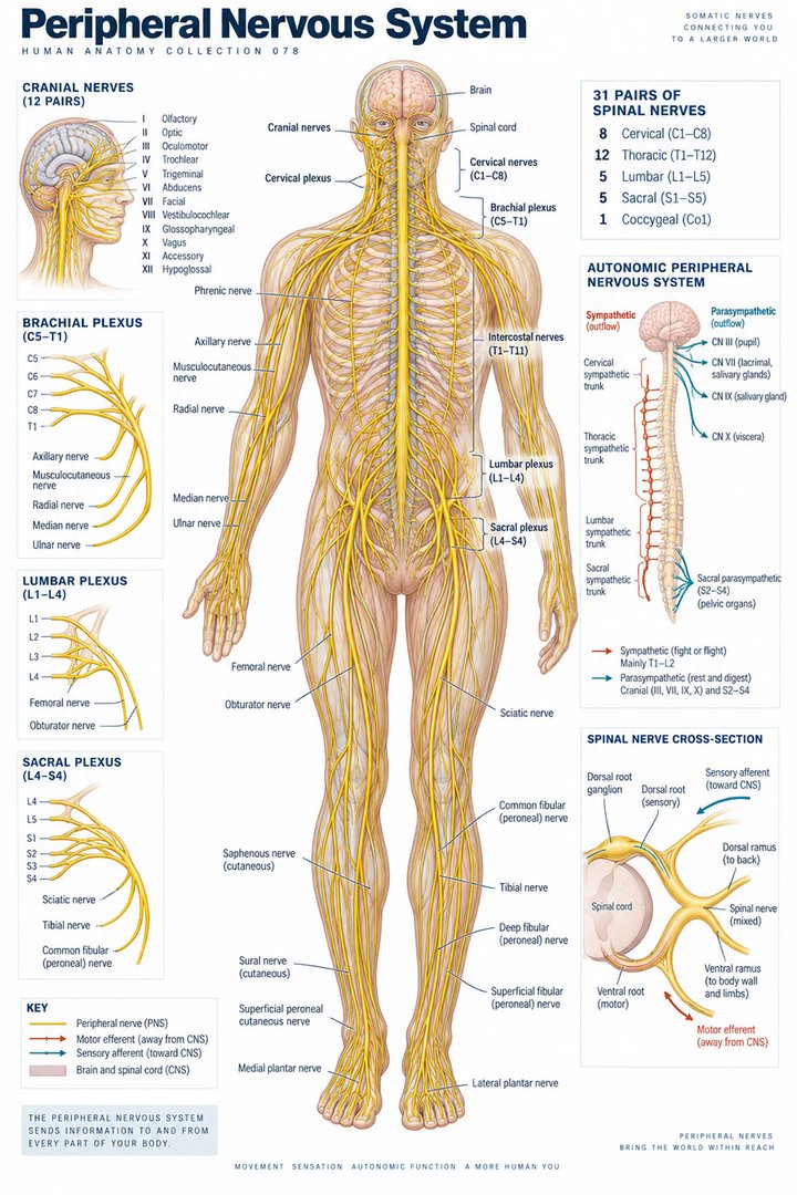 Peripheral Nervous System ↗
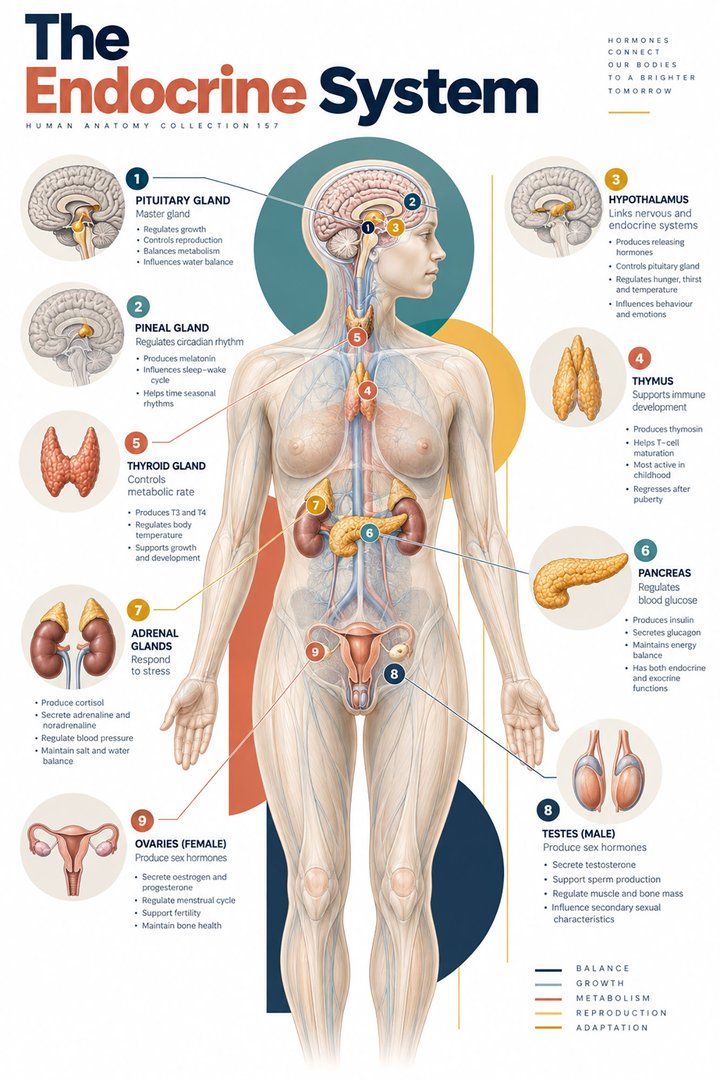 Human Endocrine Glands ↗Human Bone Density ↗Human Blood Circulation ↗
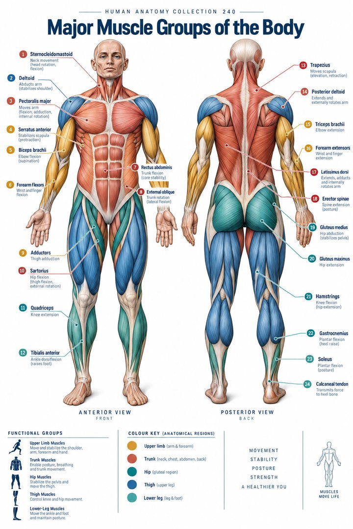 Human Muscle Groups ↗
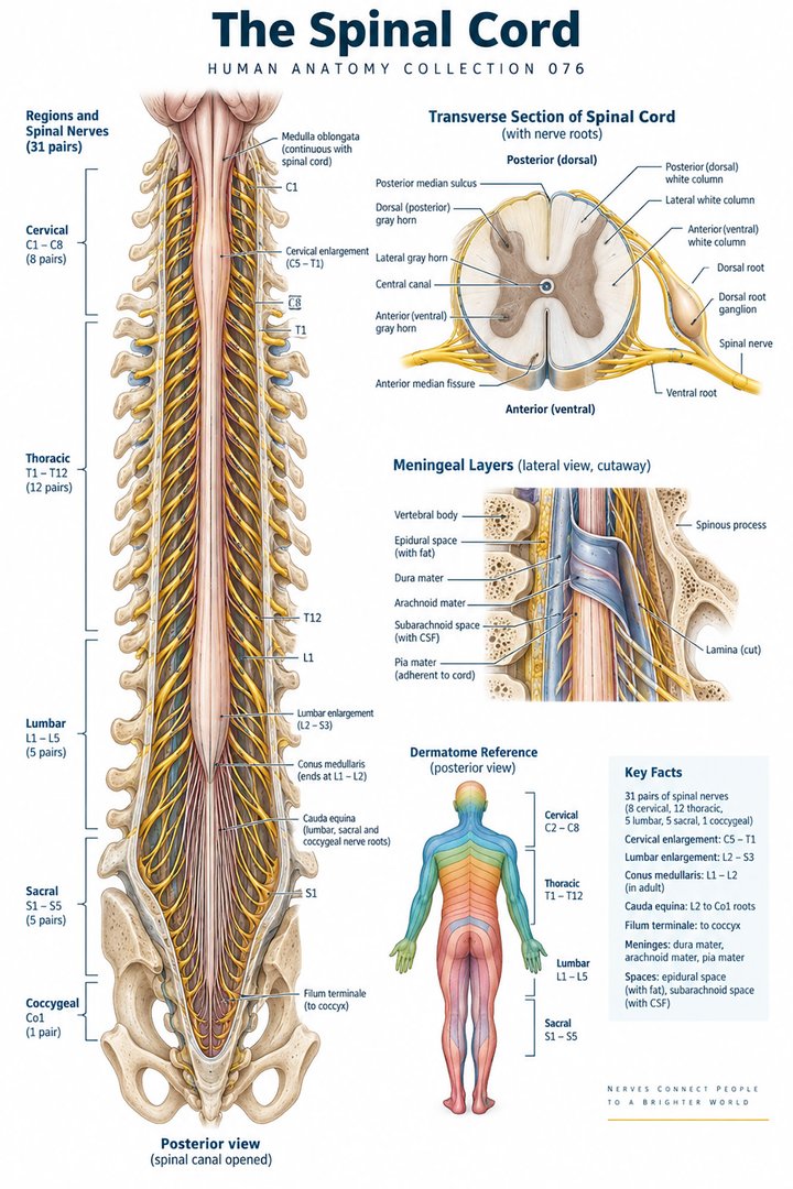 Human Spinal Cord ↗
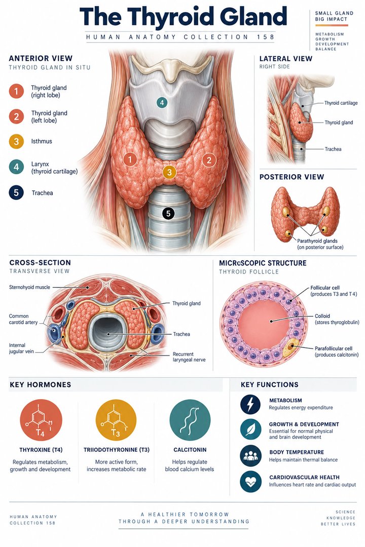 Human Thyroid Gland ↗
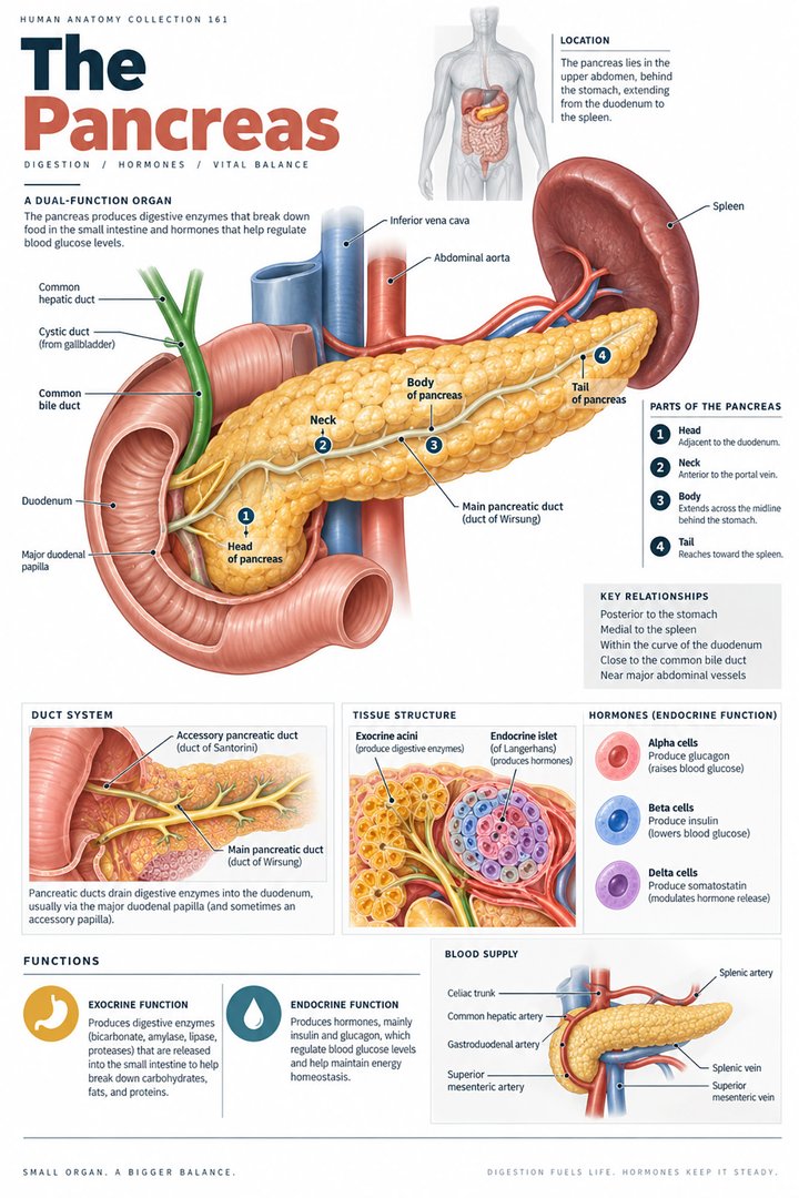 Human Pancreas Function ↗
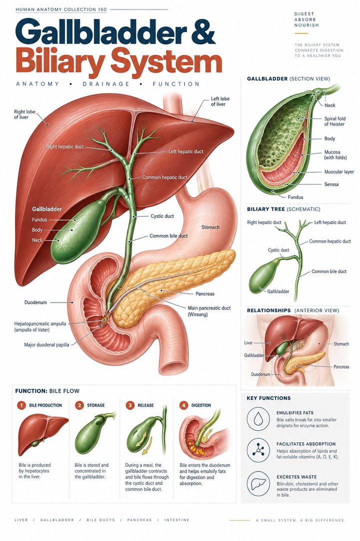 Human Gallbladder Anatomy ↗