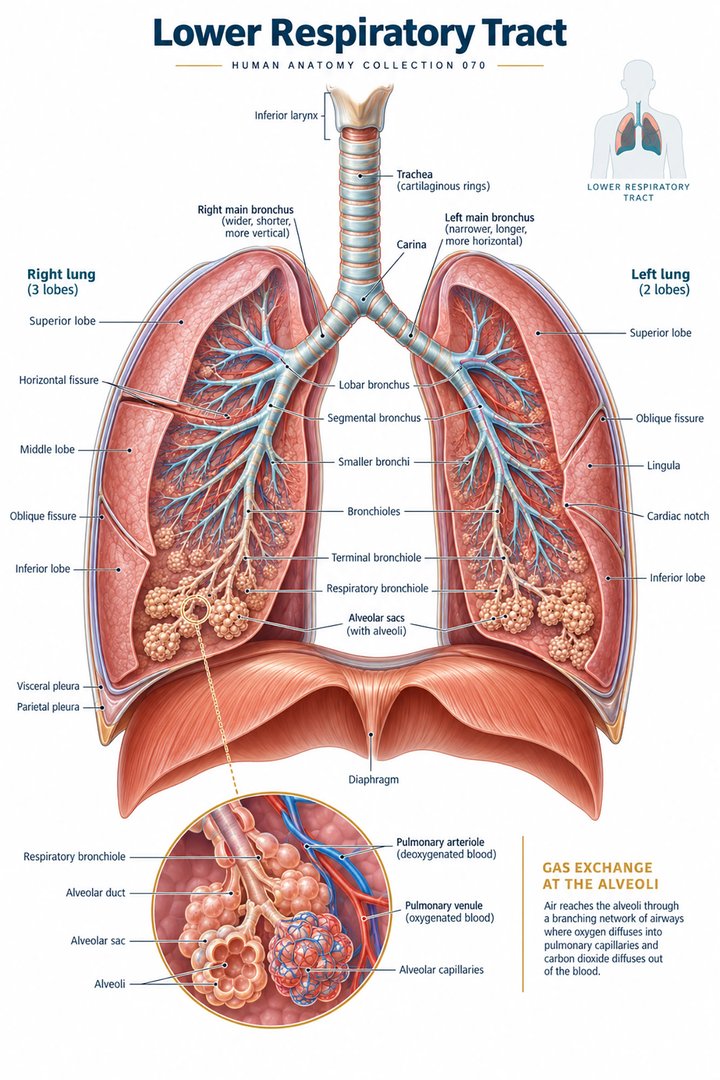
Lower Respiratory Tract
Individual poster · 300 DPI print files
Regular $9.90
Explore poster ↗$4.90USD
Offer price · Save $5.00Individual poster · 300 DPI print files
Reduced web preview. Separate high-resolution print files are supplied with the digital download.
ANATOMY 070
A modern anatomy illustration exploring lower respiratory tract, designed for educational and decorative use.
Purchases are not available yet. Checkout and download delivery are being prepared.
Sold by Kunstolymp.de · Germany
Compare prices & the complete collection ↗This is a DIGITAL DOWNLOAD. No physical product will be shipped. After purchase, download your files and print them at home, at a local print shop, or through an online professional printing service.
The separate 300 DPI print files are ideal for 24×36-inch posters (approximately 60×90 cm). ISO and 5×7 formats are included. The white border lets you adjust the layout or trim to fit other frame sizes; check the preview before printing to keep the illustration and labels visible.
The planned bundle delivery includes a PDF information sheet with a link to the relevant Google Drive folder and download instructions. The store is currently in preview and purchases are not yet available.
A science-inspired choice for a student’s study corner, an educator’s classroom, or an anatomy enthusiast’s wall. Final licensing terms will apply.
CLINICAL CONTEXT
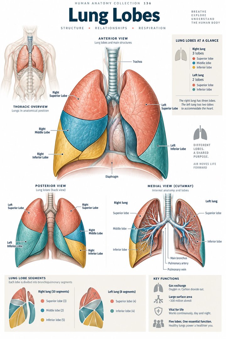
The lower respiratory tract comprises the trachea, bronchi, bronchioles, and lungs, including alveoli where oxygen and carbon dioxide exchange across the alveolar-capillary membrane. Conducting airways distribute air; elastic lung tissue and respiratory muscles support ventilation; pulmonary vessels perfuse gas-exchanging surfaces. Inflammation, obstruction, fluid, infection, or loss of elastic recoil can impair ventilation, oxygenation, or both. This anatomy helps clinicians interpret cough, sputum, wheeze, chest tightness, pleuritic pain, hypoxaemia, or breathlessness while considering age, smoking and occupational exposure, immune status, aspiration risk, medications, baseline function, and disease trajectory.
Acute bronchitis is usually viral and produces cough, sometimes mucus, chest discomfort, mild fever, or transient wheeze; cough may outlast other symptoms. Pneumonia involves infection and inflammation of lung tissue and may cause fever, tachypnoea, focal crackles, pleuritic pain, hypoxaemia, or delirium, especially in older or frail adults. Asthma causes variable airway inflammation and bronchoconstriction, while COPD commonly involves persistent airflow limitation associated with chronic bronchitis or emphysema. Exacerbations can sharply limit activity and sleep. Pulmonary embolism, pneumothorax, heart failure, lung cancer, interstitial lung disease, and medication effects may mimic or coexist with infection. Ask about onset, exertion, sputum, haemoptysis, fever, chest pain, inhaler technique, prior exacerbations, exposure, smoking, travel, immobility, and daily function.
Start with work of breathing, respiratory rate, oxygen saturation, haemodynamic stability, mental status, hydration, and ability to speak. Auscultation and percussion can guide but do not independently exclude serious disease. Chest radiography is useful when pneumonia, complication, or another diagnosis is suspected; spirometry confirms persistent airflow obstruction in stable patients rather than during an acute episode. Microbiology, blood tests, blood gases, ECG, or CT should be selected according to severity and the question being asked. Management depends on cause: supportive care for uncomplicated viral bronchitis; appropriate antibiotics for bacterial pneumonia; inhaled bronchodilators and anti-inflammatory therapy for asthma or COPD according to a clinician’s plan; oxygen for documented hypoxaemia; and escalation to hospital or ventilatory support for severe respiratory failure. Pulmonary rehabilitation, vaccination, smoking cessation, and exposure reduction can improve health and reduce future risk.
Breathing retraining, supervised exercise, relaxation, and nutrition support can complement prescribed care, particularly in chronic disease, but cannot reverse severe airflow obstruction or treat pneumonia or embolism. Steam inhalation can cause burns; herbal preparations and supplements may interact with anticoagulants, inhalers, or other medicines. Patients should discuss these approaches with a clinician and avoid delaying necessary treatment. Emergency assessment is required for severe breathlessness, inability to speak in sentences, blue or grey lips, confusion, collapse, sudden chest pain, coughing blood, rapidly falling oxygen levels, or poor response to a rescue plan. This page supports learning and does not replace local protocols or individual assessment.
Further reading: MedlinePlus: acute bronchitis · MedlinePlus: COPD · WHO: pneumonia
Explain the working diagnosis, important alternatives, planned tests, expected course, and specific safety-netting in language the patient can use. Invite questions about treatment goals, capacity, cost, culture, and complementary therapies. Use a professional interpreter when language access could affect consent or understanding, and confirm clear arrangements for timely clinical review in writing. Document the reasoning, reassess response to treatment, and coordinate care across professions when illness affects more than one system. Teaching anatomy is most useful when it supports this partnership: accurate localisation, respectful listening, timely escalation, and a plan that remains responsive to the individual patient.
HUMAN ANATOMY COLLECTION · 001—300
A closer look at what makes us human. Discover anatomy posters for your study space, classroom, and everyday curiosity.
DIGITAL DOWNLOADS · 300 DPI PRINT FILES
EXPLORE BY SYSTEM
LEARN & EXPLORE
Detailed English guides with Latin terminology, diagrams, real images, and clinical learning connections.
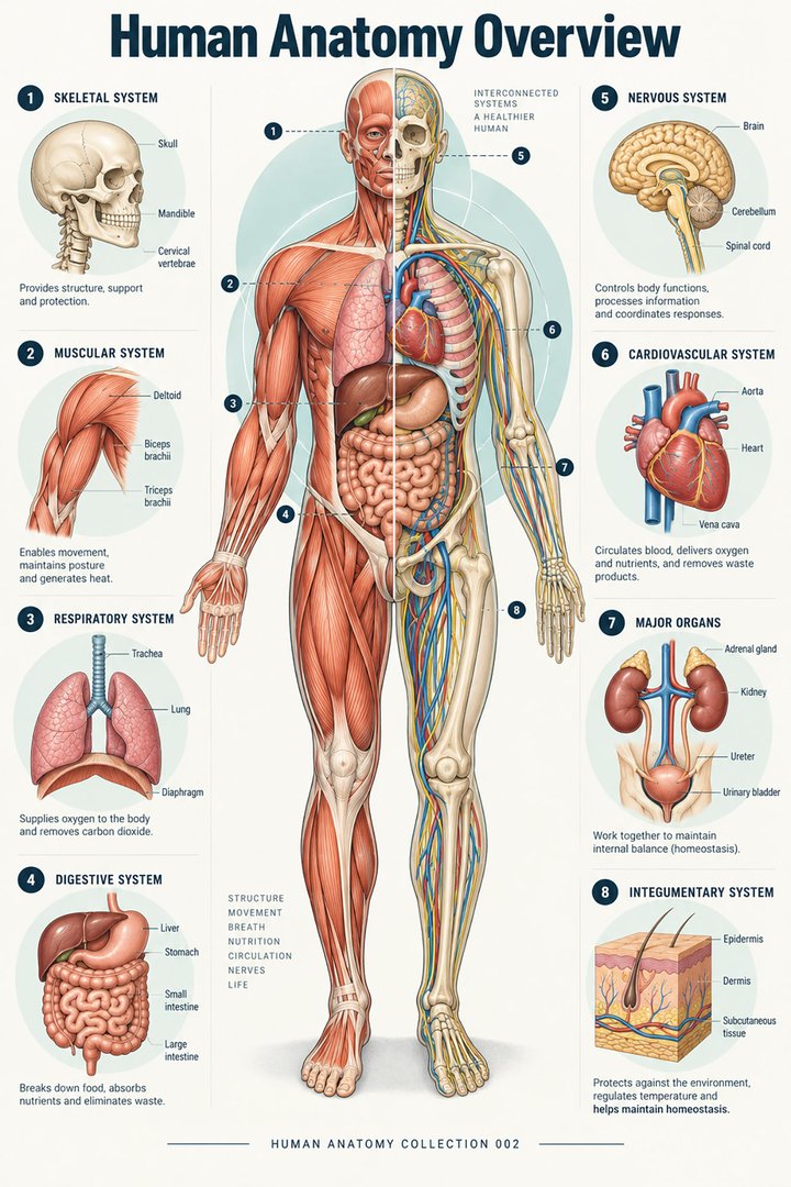 Human Anatomy Diagrams ↗
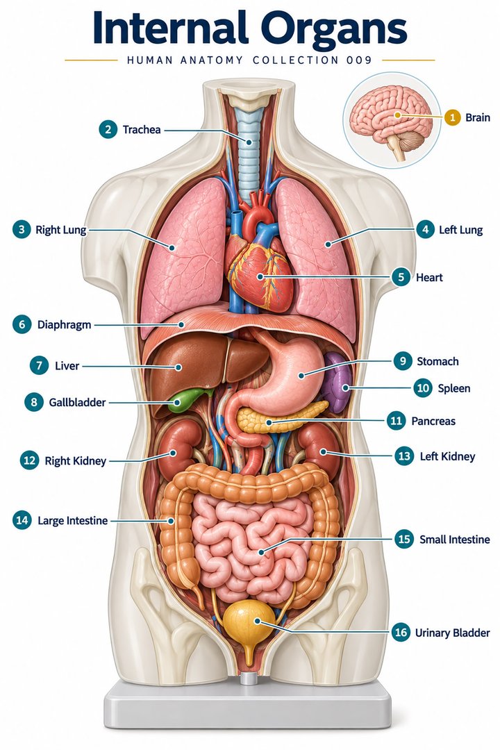 Human Body Organs ↗
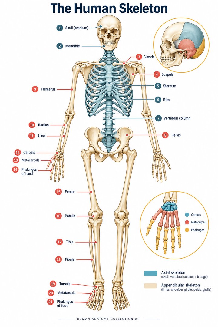 Skeletal System Anatomy ↗
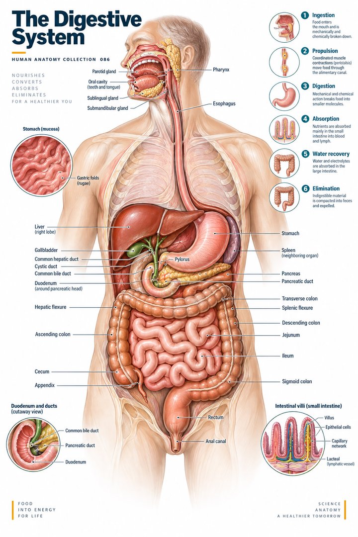 Human Digestive System ↗
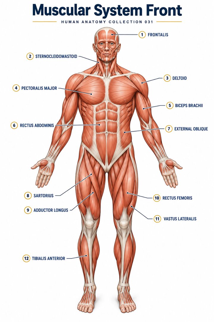 Muscular System Overview ↗
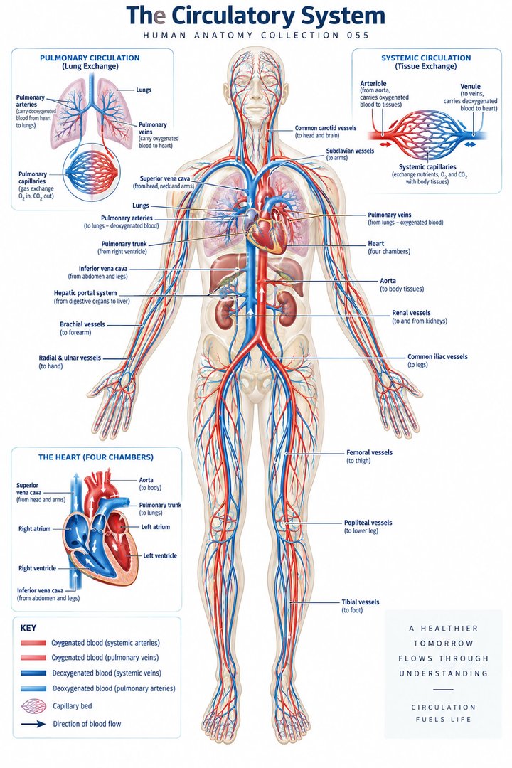 Human Circulatory System ↗
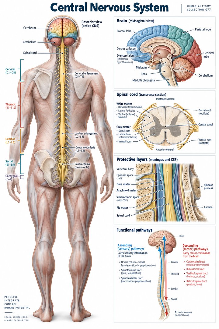 Human Nervous System ↗
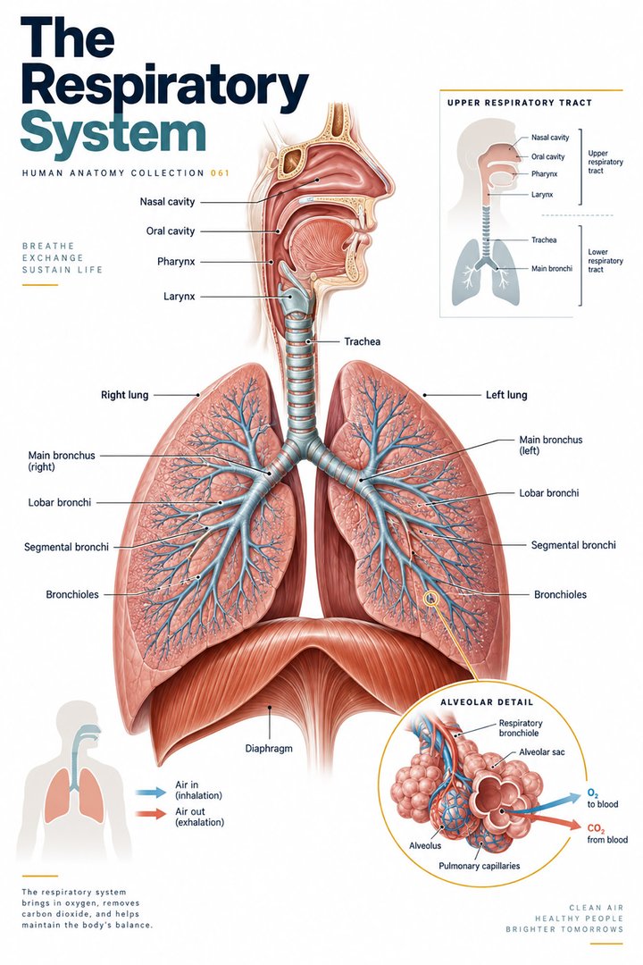 Respiratory System Functions ↗
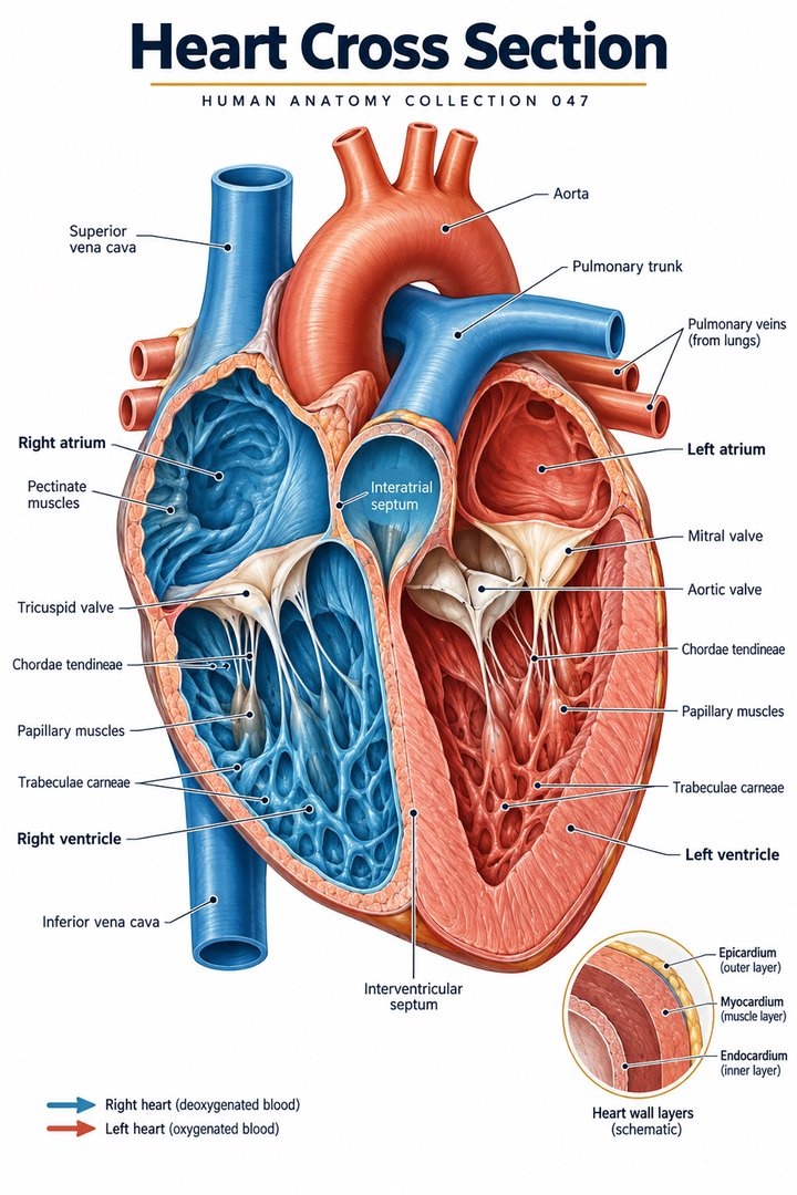 Human Heart Structure ↗
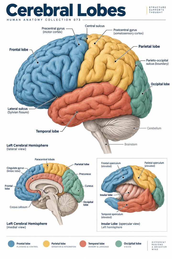 Human Brain Lobes ↗
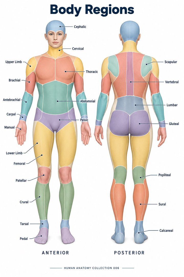 Human Body Parts ↗
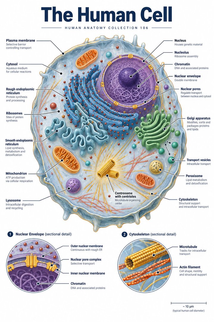 Human Cell Structure ↗
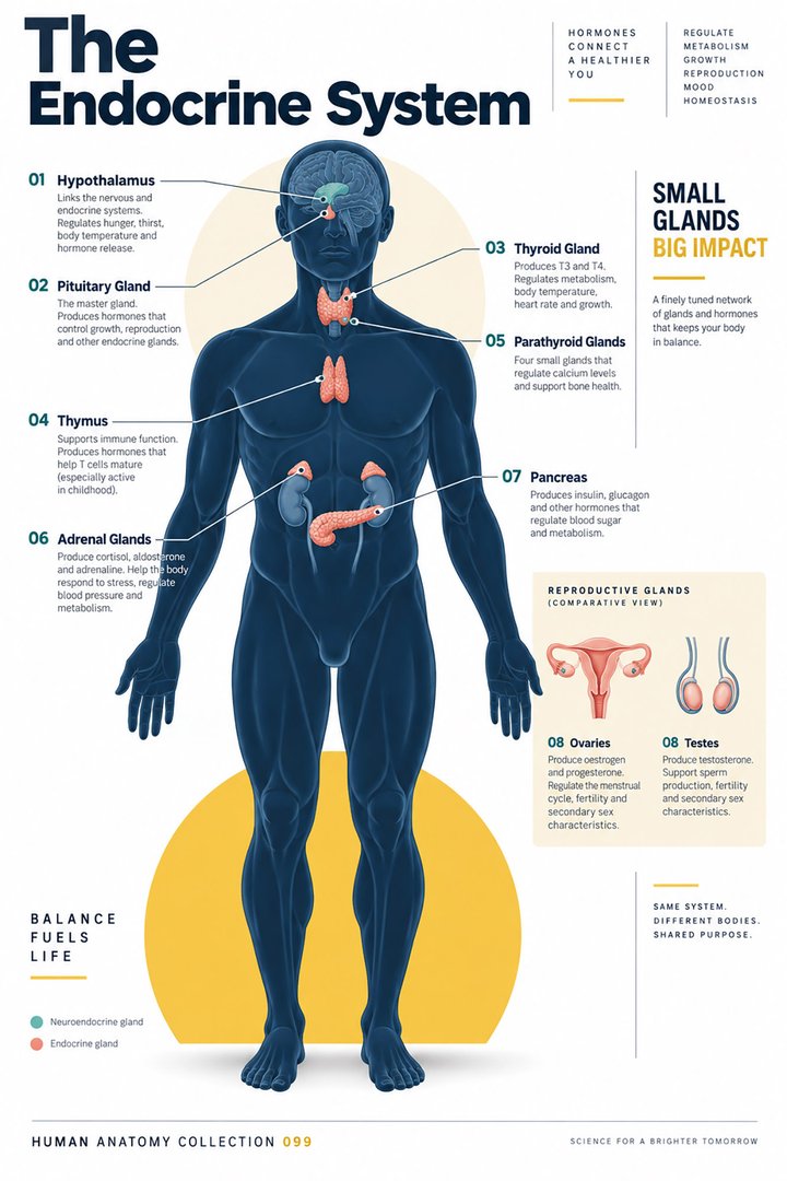 Human Endocrine System ↗
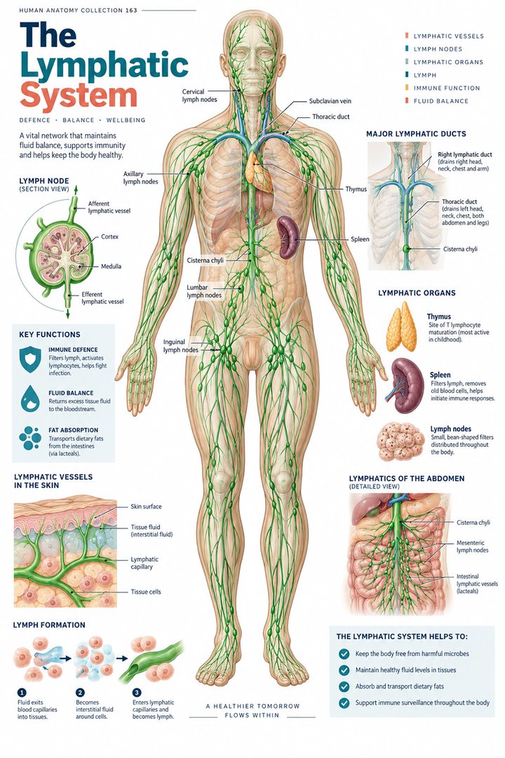 Human Lymphatic System ↗
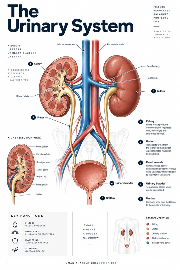 Human Urinary System ↗
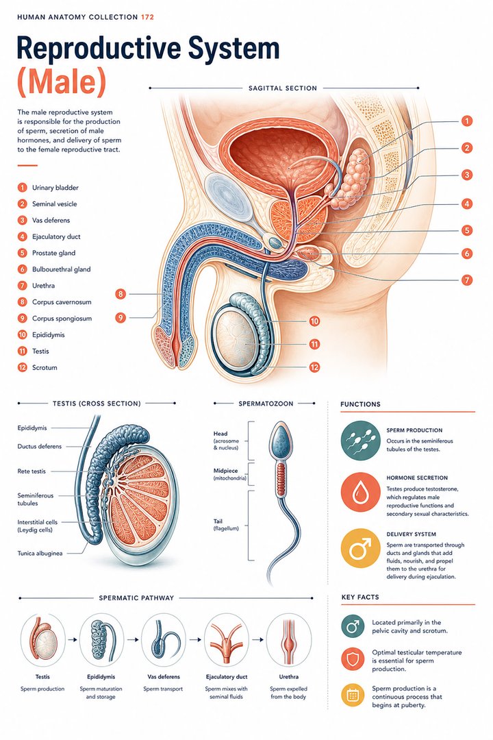 Male Reproductive System ↗
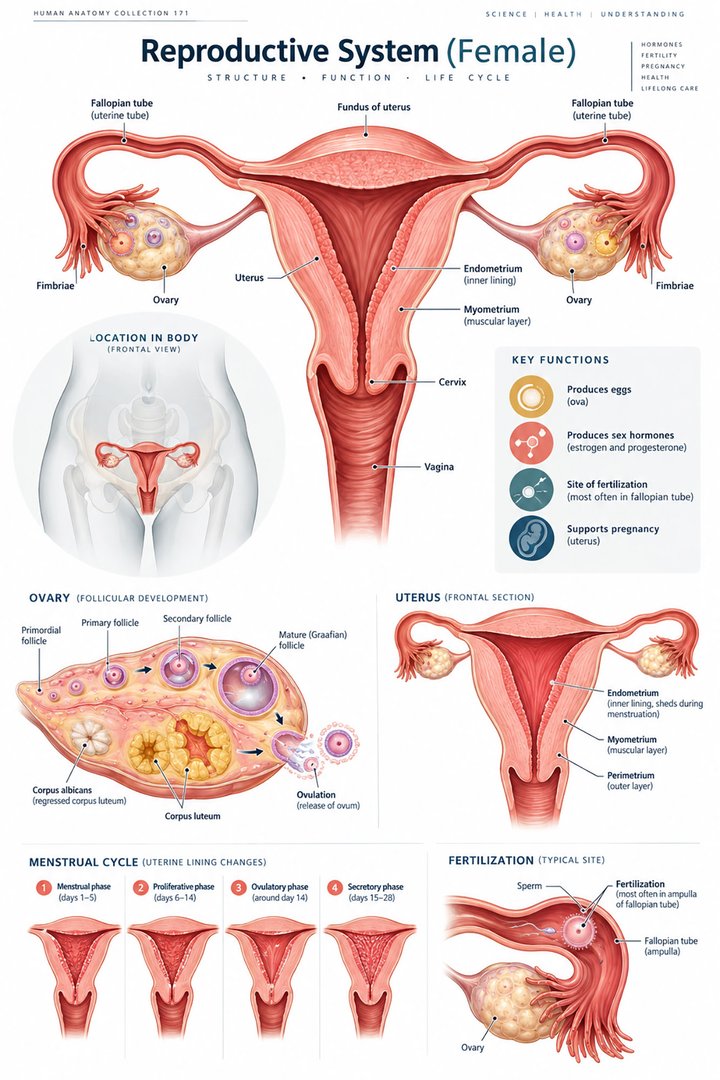 Female Reproductive System ↗
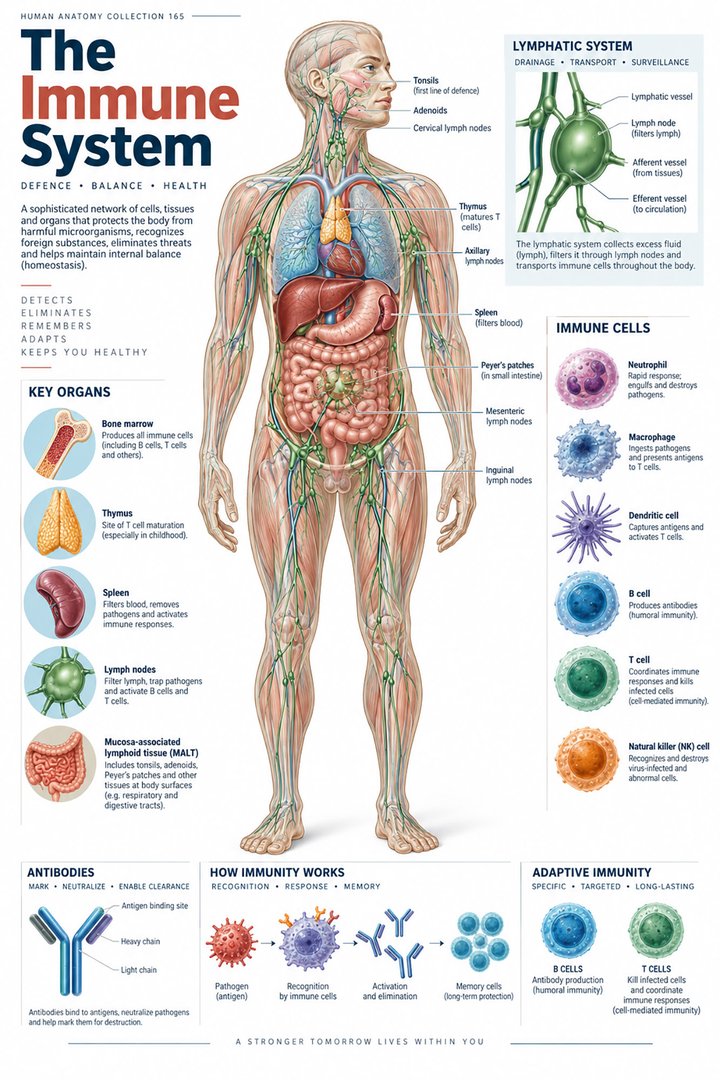 Human Immune System ↗Central Nervous System ↗
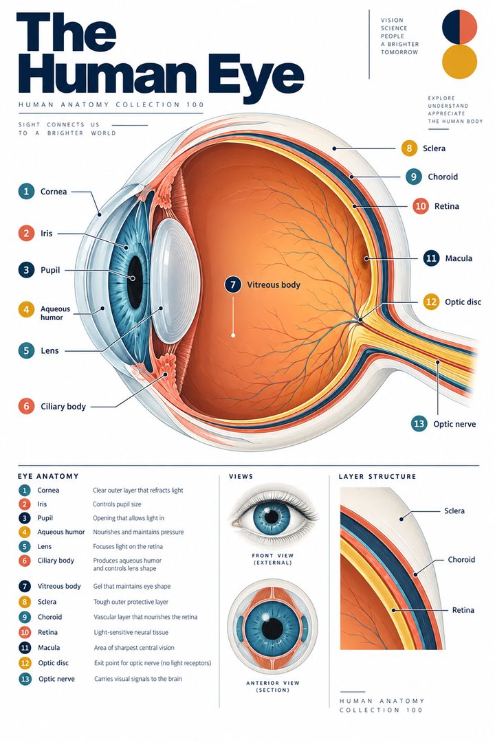 Human Eye Structure ↗
 Human Ear Structure ↗
 Human Liver Function ↗
 Human Kidney Anatomy ↗
 Human Lung Anatomy ↗
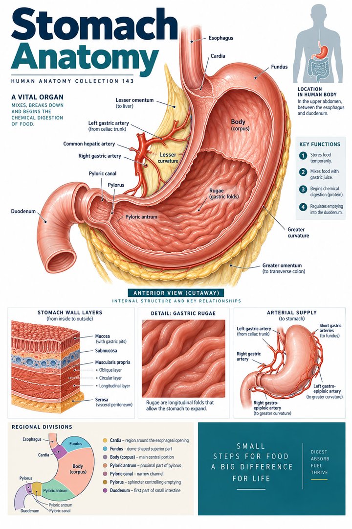 Human Stomach Structure ↗
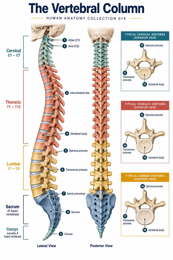 Human Spine Anatomy ↗
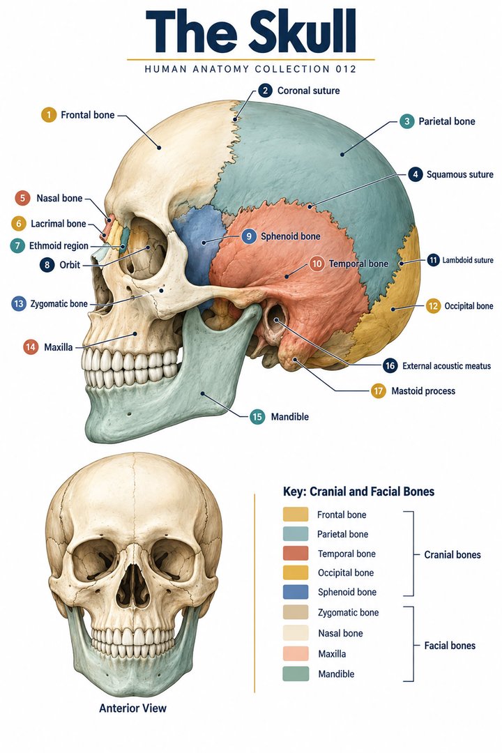 Human Skull Anatomy ↗
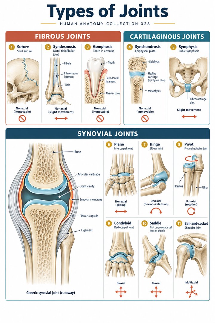 Human Joint Types ↗
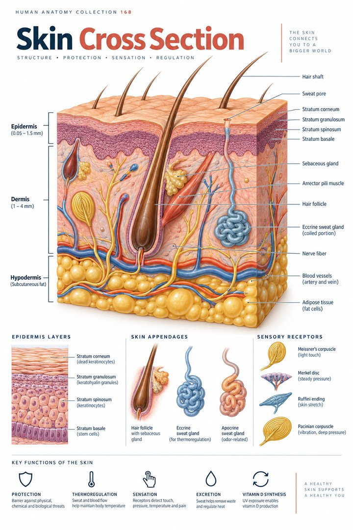 Human Skin Layers ↗
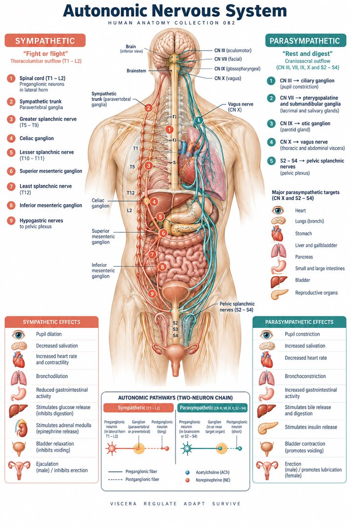 Autonomic Nervous System ↗
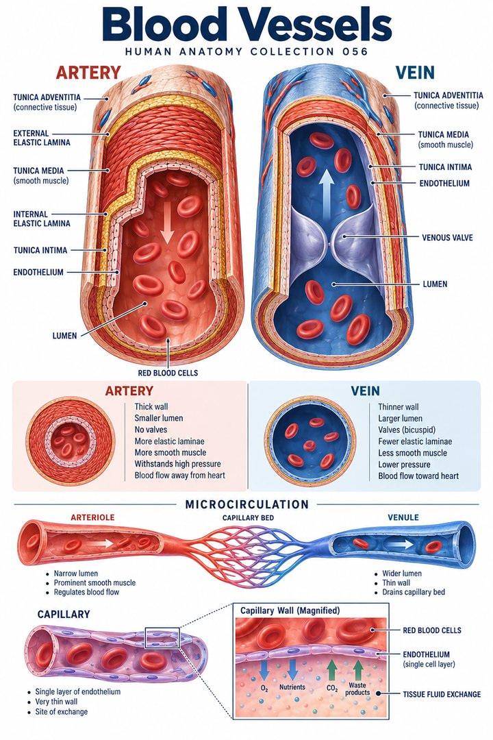 Human Blood Vessels ↗
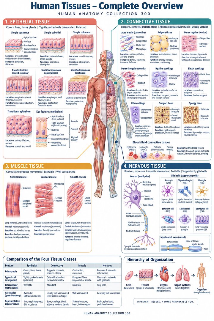 Human Body Tissues ↗
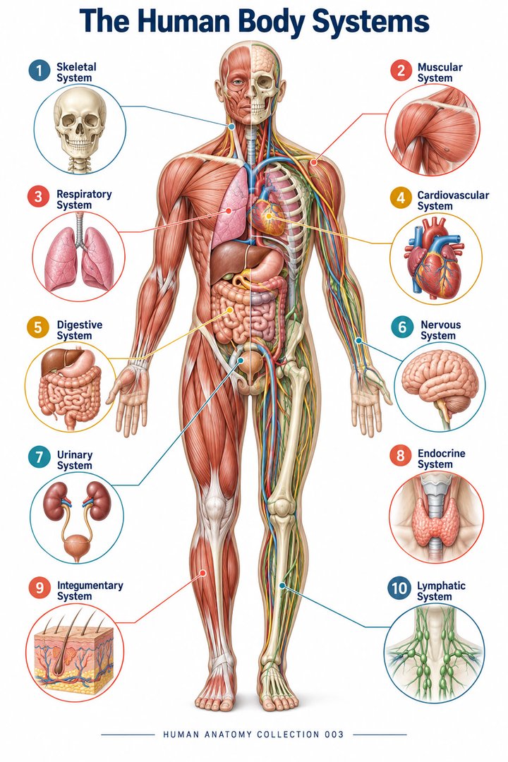 Human Body Systems ↗
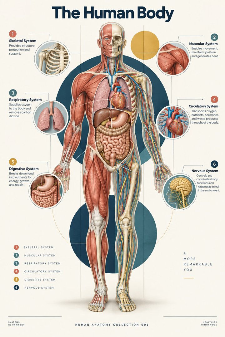 Human Body Facts ↗Human Anatomy and Physiology ↗
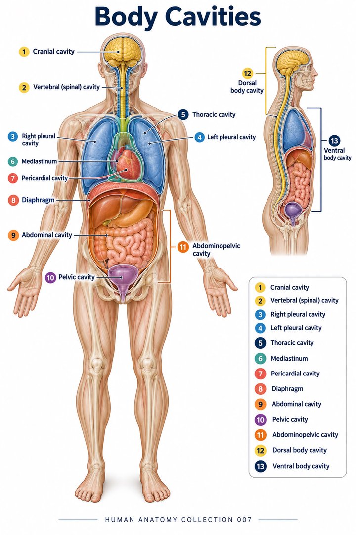 Human Body Cavities ↗Human Torso Anatomy ↗Human Digestive Tract ↗
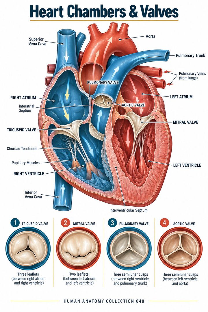 Human Heart Chambers ↗Human Organ Placement ↗
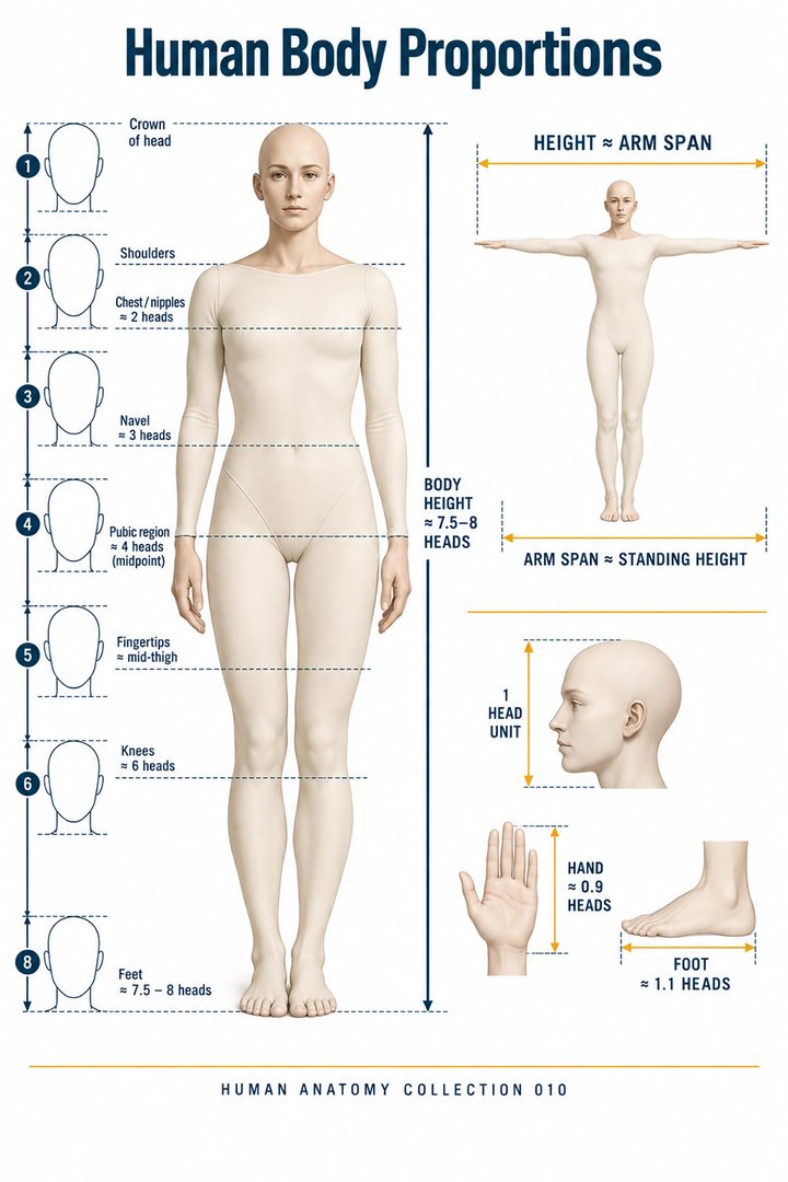 Human Body Proportions ↗
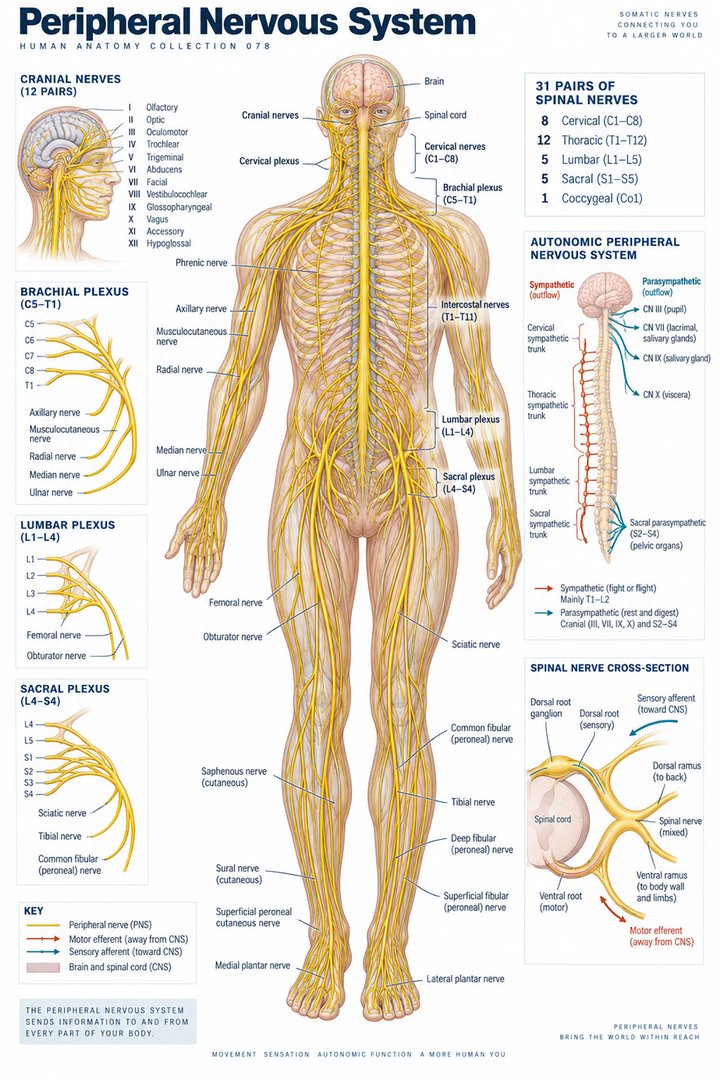 Peripheral Nervous System ↗
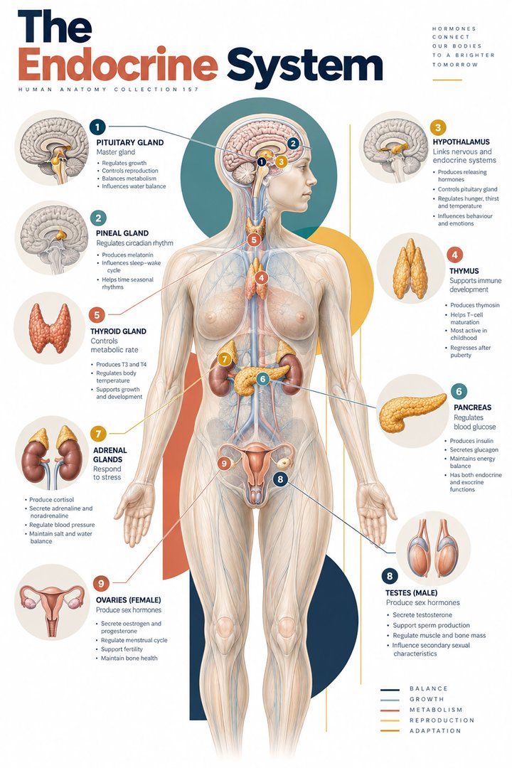 Human Endocrine Glands ↗
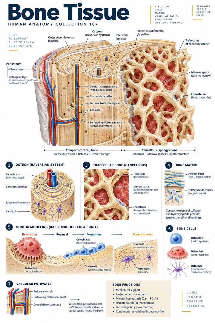 Human Bone Density ↗Human Blood Circulation ↗
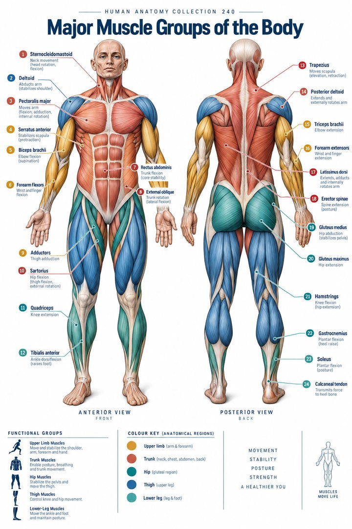 Human Muscle Groups ↗
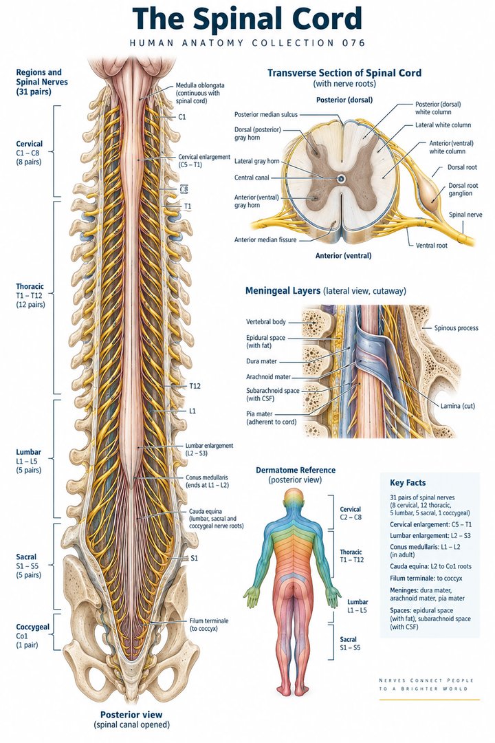 Human Spinal Cord ↗
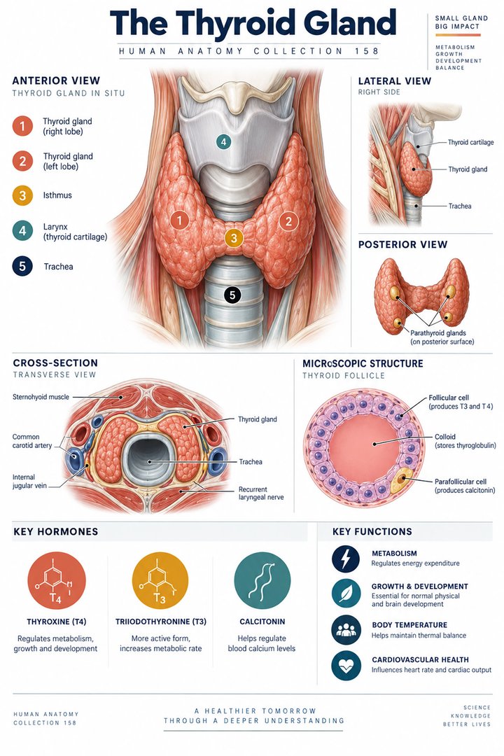 Human Thyroid Gland ↗
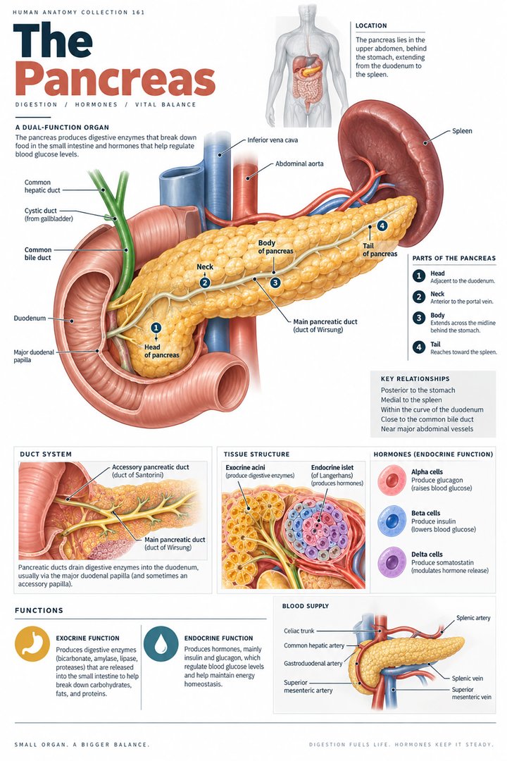 Human Pancreas Function ↗
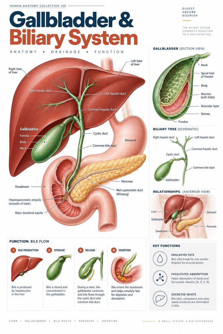 Human Gallbladder Anatomy ↗