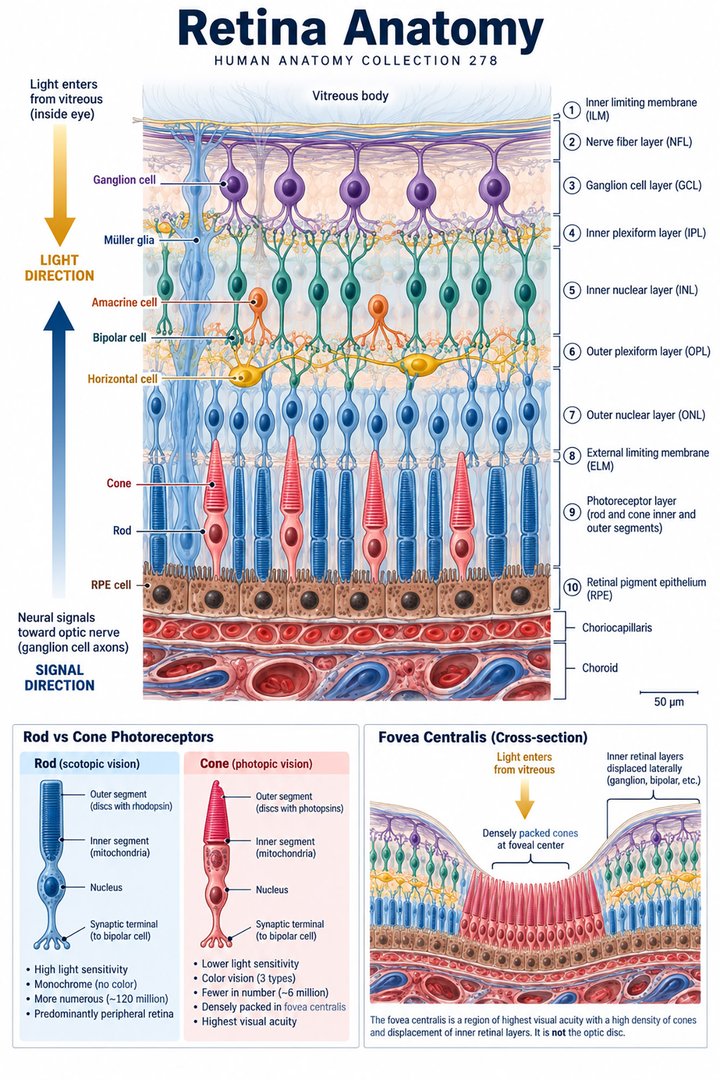
Retina Anatomy
Individual poster · 300 DPI print files
Regular $9.90
Explore poster ↗$4.90USD
Offer price · Save $5.00Individual poster · 300 DPI print files
Reduced web preview. Separate high-resolution print files are supplied with the digital download.
ANATOMY 278
A modern anatomy illustration exploring retina anatomy, designed for educational and decorative use.
Purchases are not available yet. Checkout and download delivery are being prepared.
Sold by Kunstolymp.de · Germany
Compare prices & the complete collection ↗This is a DIGITAL DOWNLOAD. No physical product will be shipped. After purchase, download your files and print them at home, at a local print shop, or through an online professional printing service.
The separate 300 DPI print files are ideal for 24×36-inch posters (approximately 60×90 cm). ISO and 5×7 formats are included. The white border lets you adjust the layout or trim to fit other frame sizes; check the preview before printing to keep the illustration and labels visible.
The planned bundle delivery includes a PDF information sheet with a link to the relevant Google Drive folder and download instructions. The store is currently in preview and purchases are not yet available.
A science-inspired choice for a student’s study corner, an educator’s classroom, or an anatomy enthusiast’s wall. Final licensing terms will apply.
CLINICAL CONTEXT
The retina is a layered neural tissue lining the back of the eye. Photoreceptors convert light into electrical signals; bipolar and horizontal cells process local input; amacrine cells shape temporal and motion signals; ganglion-cell axons form the optic nerve. The macula supports detailed central vision, with the fovea providing highest acuity. Retinal pigment epithelium supports photoreceptors and participates in outer-retinal metabolism. The inner retina receives blood primarily from retinal vessels, while the outer retina relies on the choroidal circulation. This anatomy explains why vascular leakage, ischaemia, degeneration, inflammation, or separation from supporting tissue can affect central detail, colour, night vision, or peripheral fields in different patterns.
Diabetic retinopathy follows chronic metabolic injury to retinal microvasculature. Early non-proliferative disease may be asymptomatic; later leakage can cause diabetic macular oedema, while ischaemia may stimulate fragile new vessels that bleed or scar. Age-related macular degeneration may cause gradual distortion or central blur; the neovascular form can progress quickly. A retinal tear or detachment may present with new flashes, floaters, or a curtain-like shadow and is an ophthalmic emergency. Retinal vein occlusion can cause sudden painless visual loss; arterial occlusion is also time-critical and may signal systemic vascular risk. Patients may report difficulty reading, face recognition, night driving, or seeing straight lines. Ask about onset, one or both eyes, field defects, diabetes duration and control, blood pressure, smoking, prior eye procedures, anticoagulation, trauma, and family history.
Check visual acuity and pupils, then arrange dilated fundus examination and retinal imaging such as optical coherence tomography or fundus photography when indicated. Fluorescein angiography or ocular ultrasound may help define vascular leakage, ischaemia, haemorrhage, or detachment. Do not delay urgent referral for suspected detachment, arterial occlusion, or sudden major visual loss while pursuing routine testing. Diabetic retinopathy care combines systemic risk management and scheduled retinal screening with ophthalmic treatment based on severity. Intravitreal anti-VEGF therapy is commonly used for macular oedema and neovascular AMD; laser or vitrectomy may be appropriate for selected proliferative disease, haemorrhage, traction, or detachment. Treatment aims to preserve remaining vision, reduce progression, and support rehabilitation; low-vision services, magnification, lighting changes, and occupational support can maintain independence.
Smoking cessation, blood pressure and glucose management, nutritious food, and keeping retinal appointments support overall risk reduction. AREDS2 supplements can lower progression risk in eligible people with intermediate or advanced AMD, but do not prevent AMD in healthy eyes or replace injections and monitoring. They are not suitable for everyone, so discuss formulation and interactions with a clinician. No eye exercise, detox, herbal drop, or supplement can reattach a detached retina or reverse established vascular damage. Avoid unregulated products and any delay in specialist treatment. New flashes with many floaters, a curtain or shadow, sudden painless visual loss, rapidly worsening distortion, or a painful red eye requires urgent same-day ophthalmic or emergency assessment. This article is educational only.
Further reading: MedlinePlus: retinal disorders · NEI: diabetic eye disease · Moorfields: retinal detachment
Explain the working diagnosis, important alternatives, planned tests, expected course, and specific safety-netting in language the patient can use. Invite questions about treatment goals, capacity, cost, culture, and complementary therapies. Use a professional interpreter when language access could affect consent or understanding, and confirm clear arrangements for timely clinical review in writing. Document the reasoning, reassess response to treatment, and coordinate care across professions when illness affects more than one system. Teaching anatomy is most useful when it supports this partnership: accurate localisation, respectful listening, timely escalation, and a plan that remains responsive to the individual patient.
HUMAN ANATOMY COLLECTION · 001—300
A closer look at what makes us human. Discover anatomy posters for your study space, classroom, and everyday curiosity.
DIGITAL DOWNLOADS · 300 DPI PRINT FILES
EXPLORE BY SYSTEM
LEARN & EXPLORE
Detailed English guides with Latin terminology, diagrams, real images, and clinical learning connections.
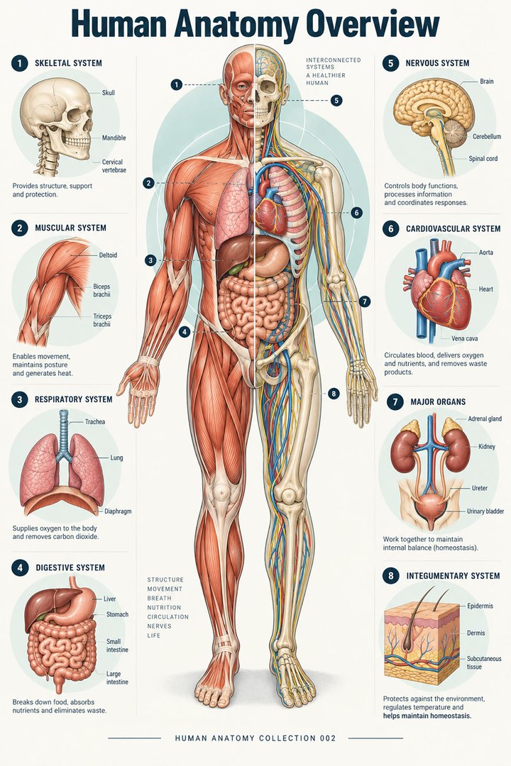 Human Anatomy Diagrams ↗
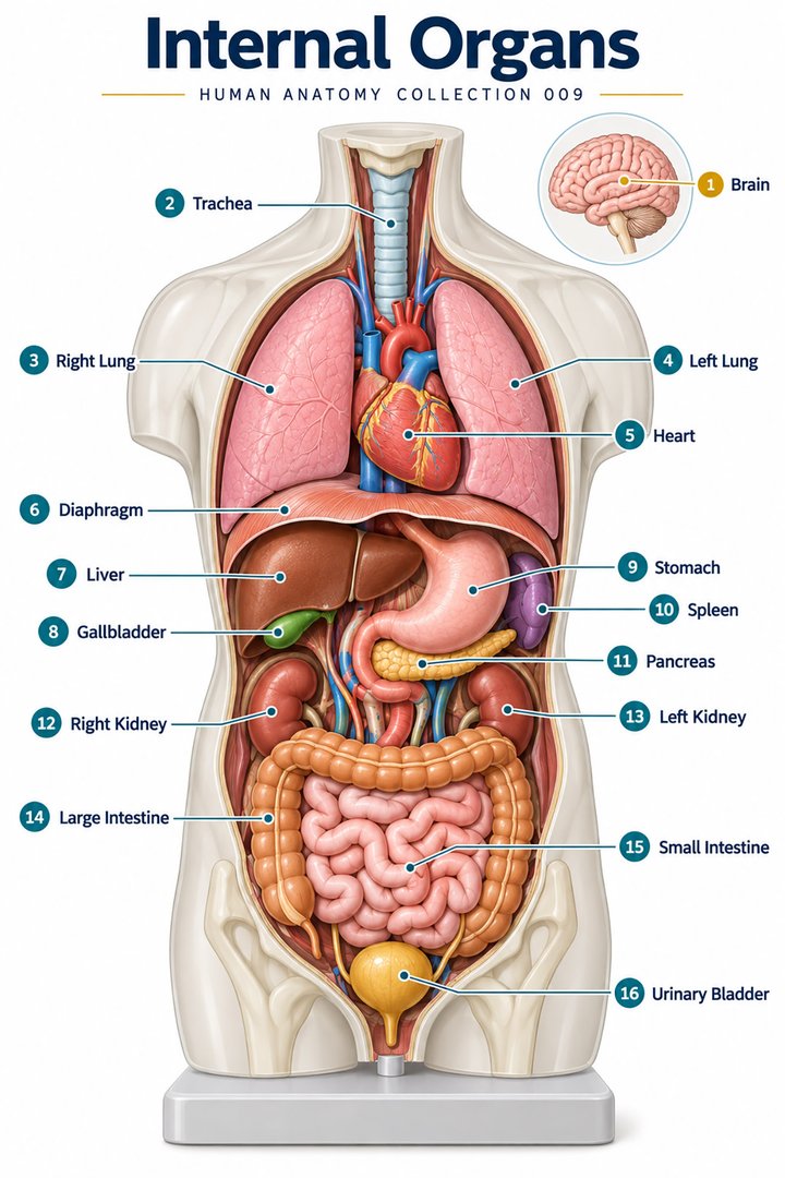 Human Body Organs ↗
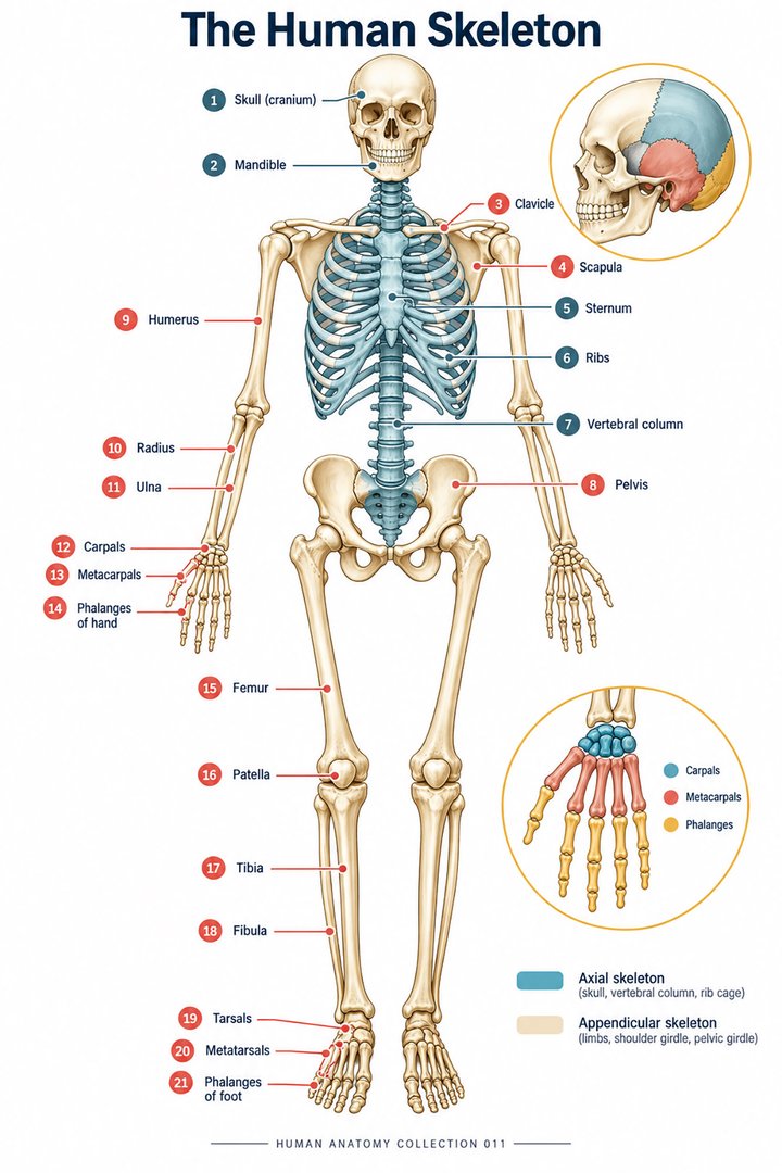 Skeletal System Anatomy ↗
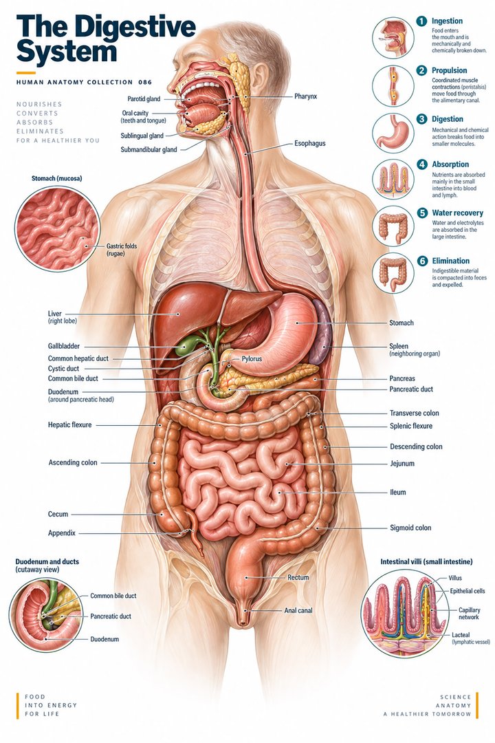 Human Digestive System ↗
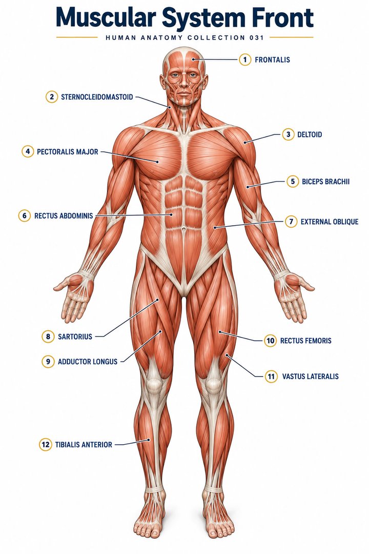 Muscular System Overview ↗
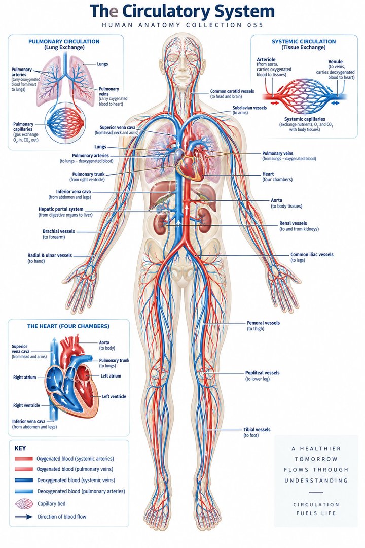 Human Circulatory System ↗
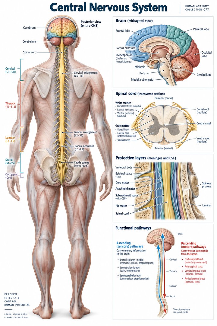 Human Nervous System ↗
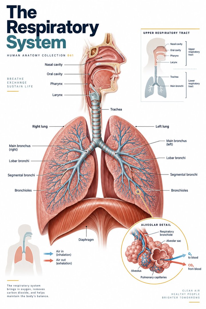 Respiratory System Functions ↗
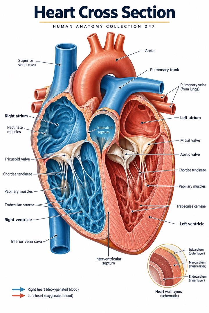 Human Heart Structure ↗
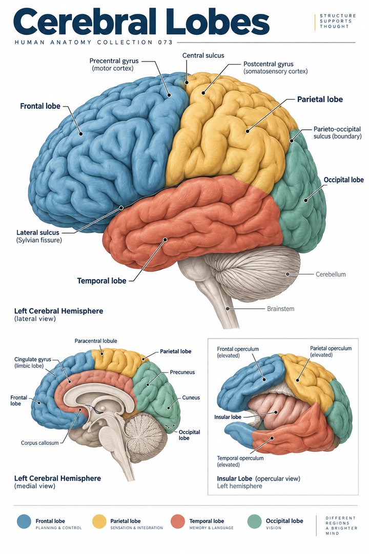 Human Brain Lobes ↗
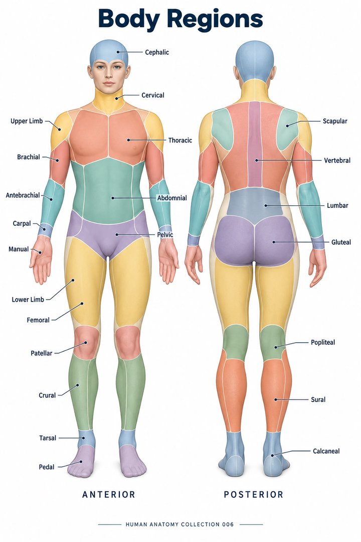 Human Body Parts ↗
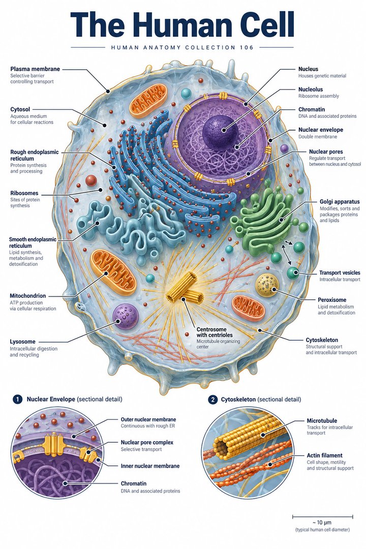 Human Cell Structure ↗
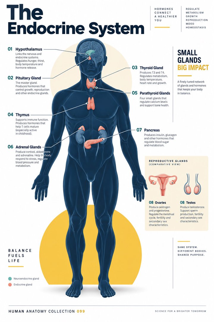 Human Endocrine System ↗
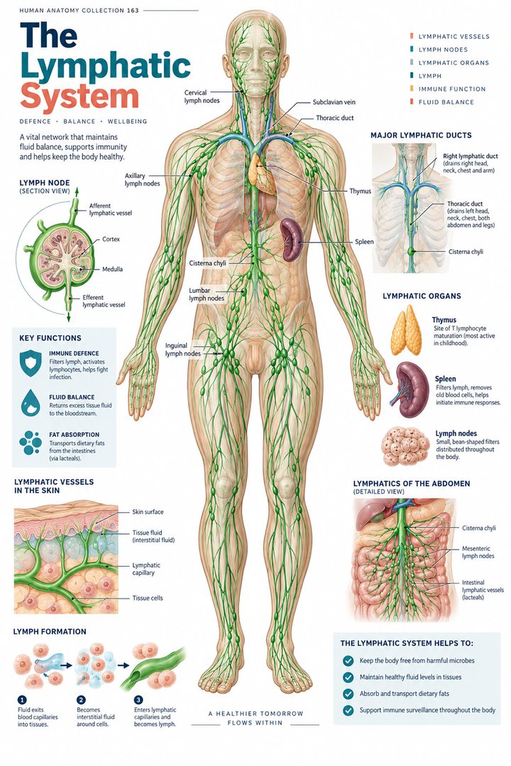 Human Lymphatic System ↗
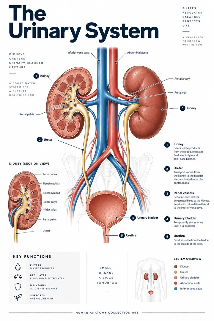 Human Urinary System ↗
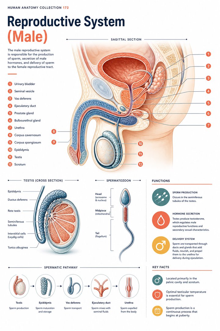 Male Reproductive System ↗
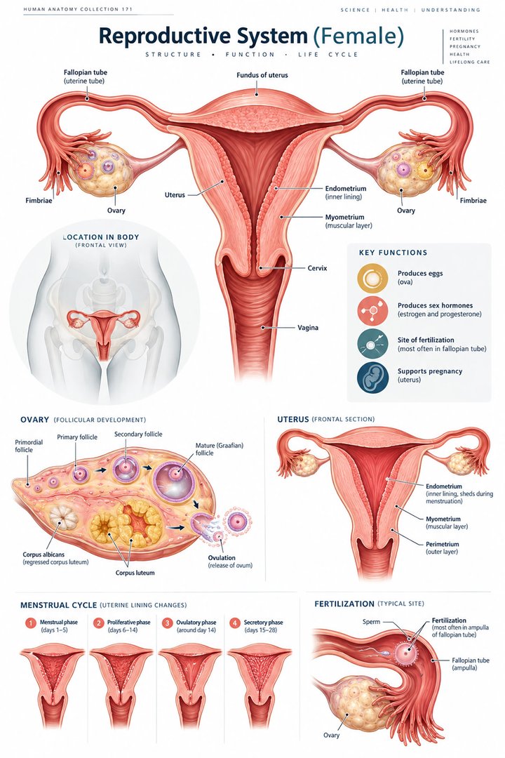 Female Reproductive System ↗
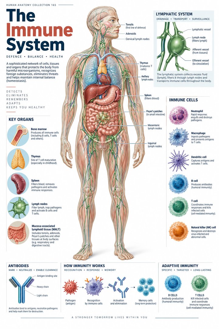 Human Immune System ↗Central Nervous System ↗
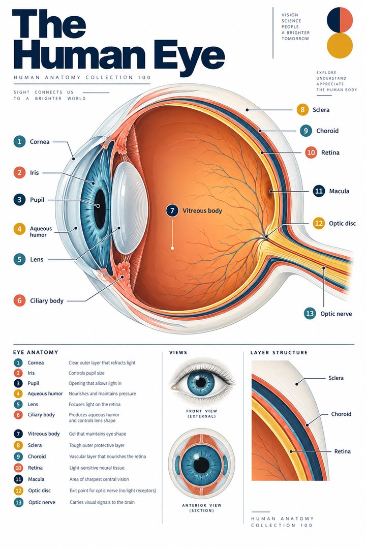 Human Eye Structure ↗
 Human Ear Structure ↗
 Human Liver Function ↗
 Human Kidney Anatomy ↗
 Human Lung Anatomy ↗
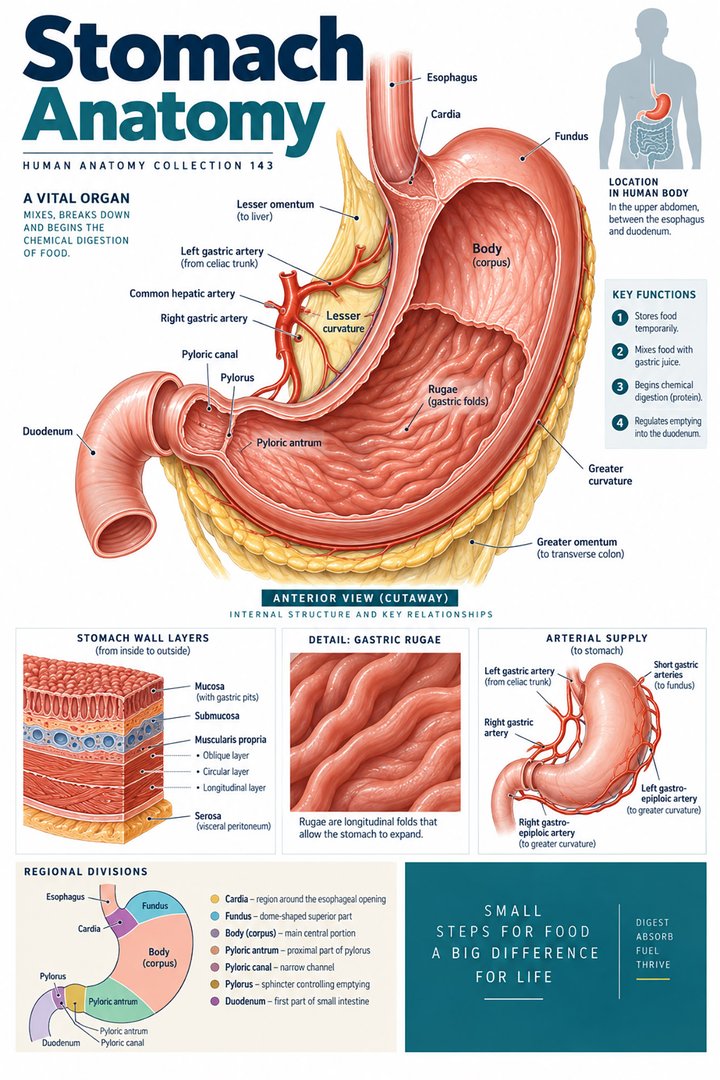 Human Stomach Structure ↗
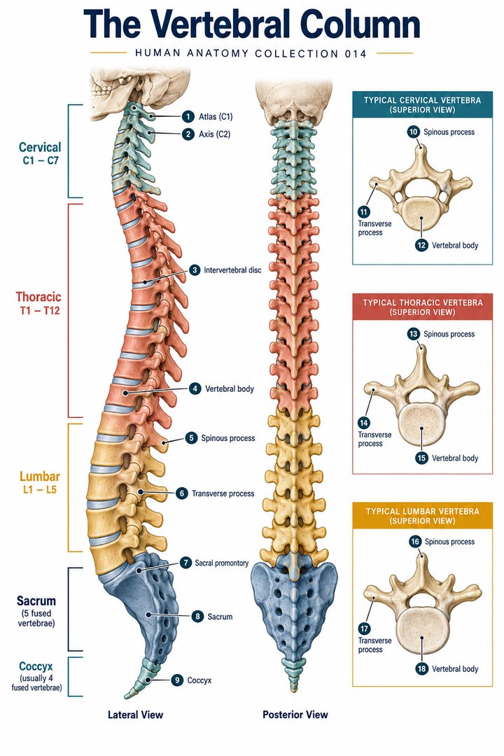 Human Spine Anatomy ↗
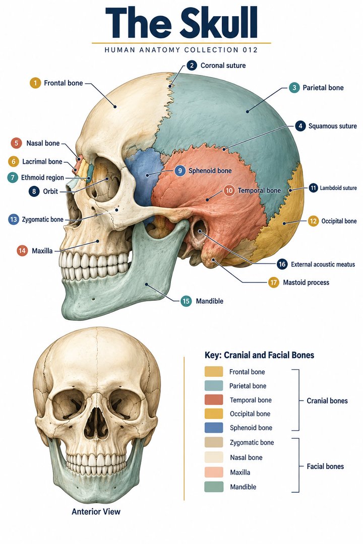 Human Skull Anatomy ↗
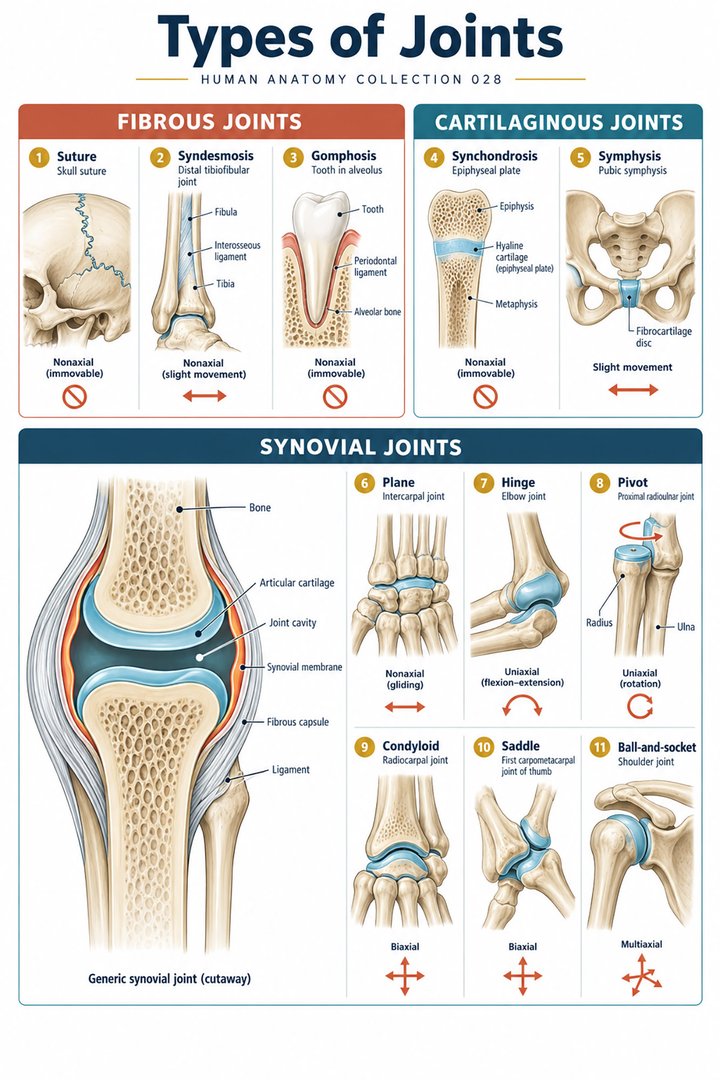 Human Joint Types ↗
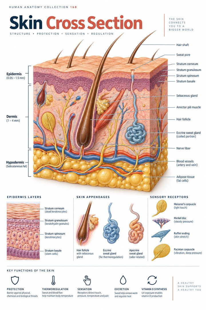 Human Skin Layers ↗
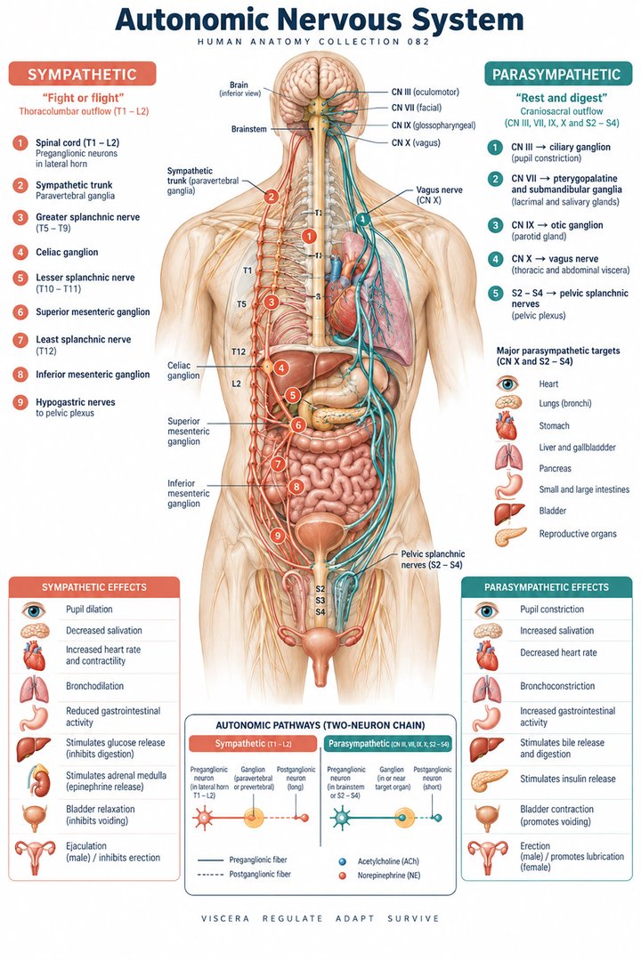 Autonomic Nervous System ↗
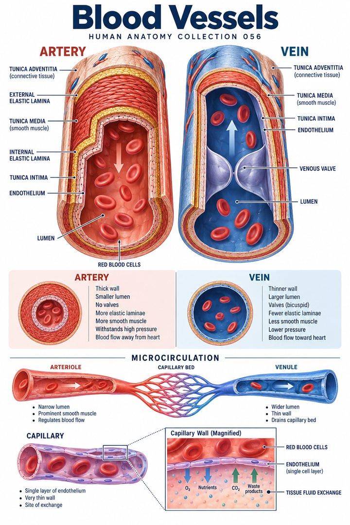 Human Blood Vessels ↗
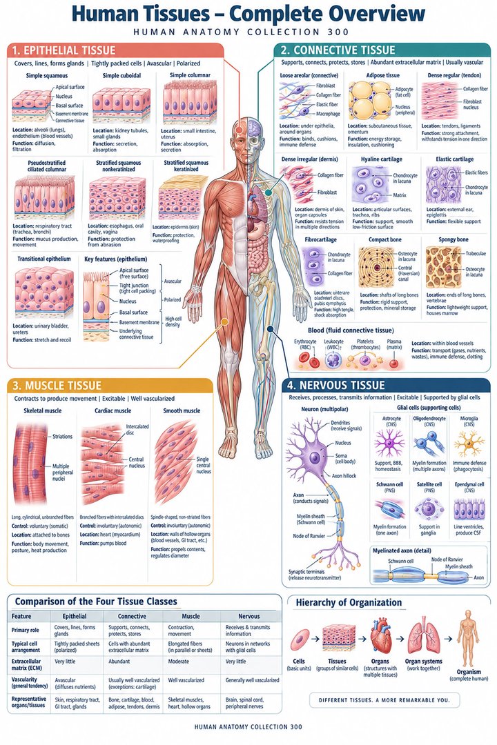 Human Body Tissues ↗
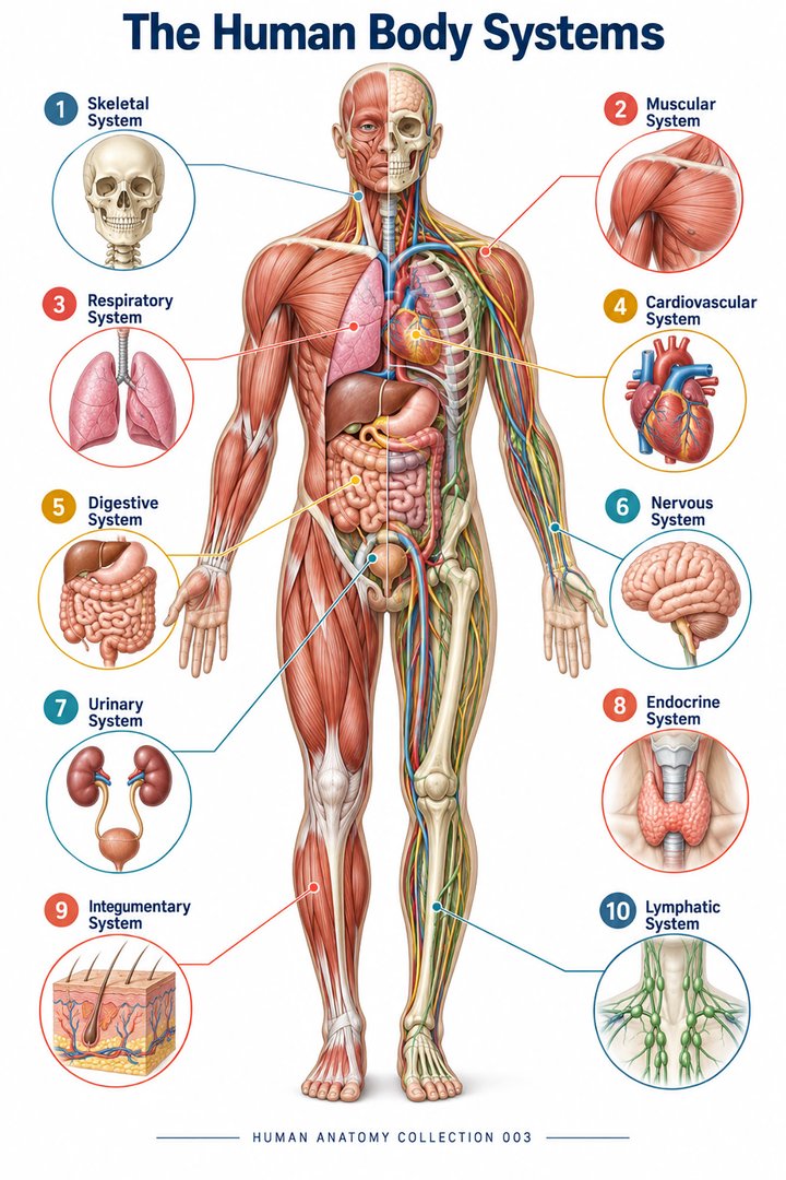 Human Body Systems ↗
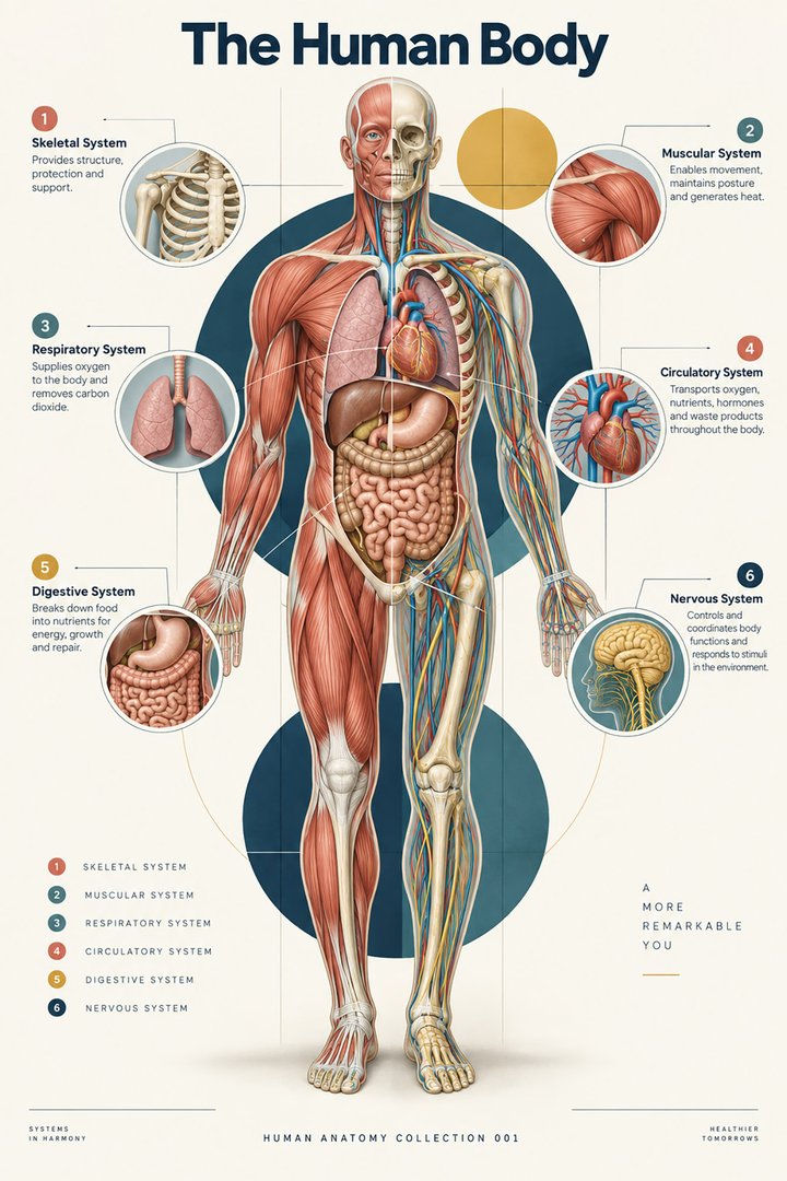 Human Body Facts ↗Human Anatomy and Physiology ↗
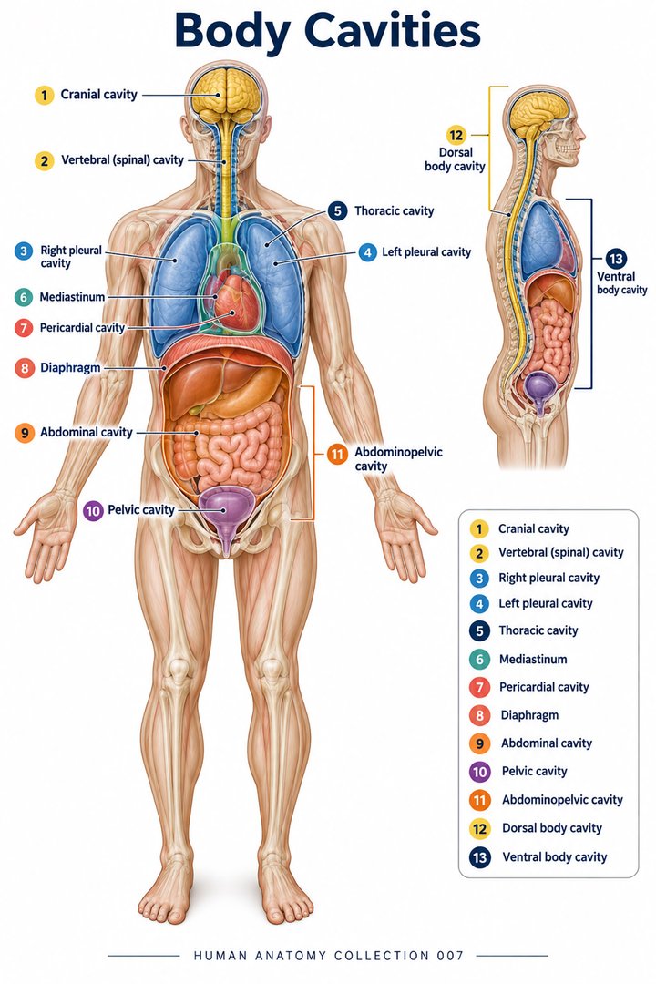 Human Body Cavities ↗Human Torso Anatomy ↗Human Digestive Tract ↗
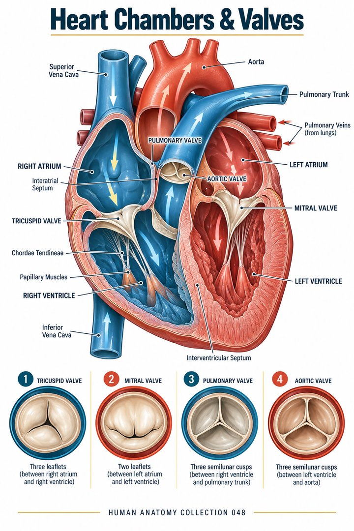 Human Heart Chambers ↗Human Organ Placement ↗
 Human Body Proportions ↗
 Peripheral Nervous System ↗
 Human Endocrine Glands ↗
 Human Bone Density ↗Human Blood Circulation ↗
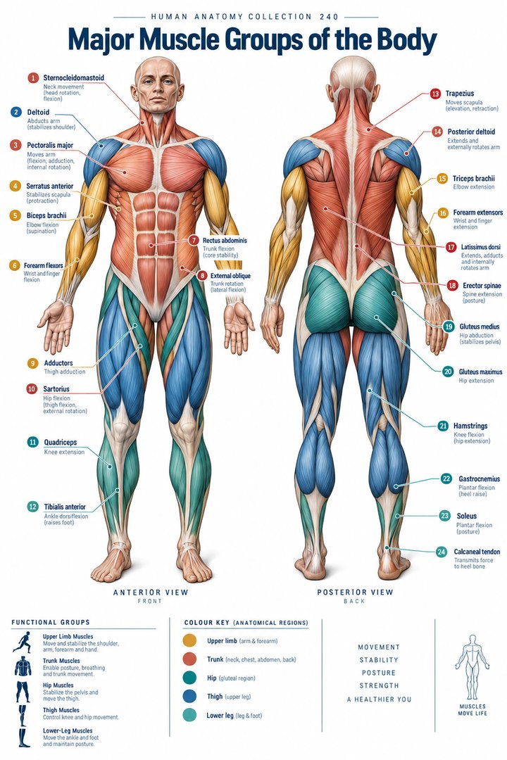 Human Muscle Groups ↗
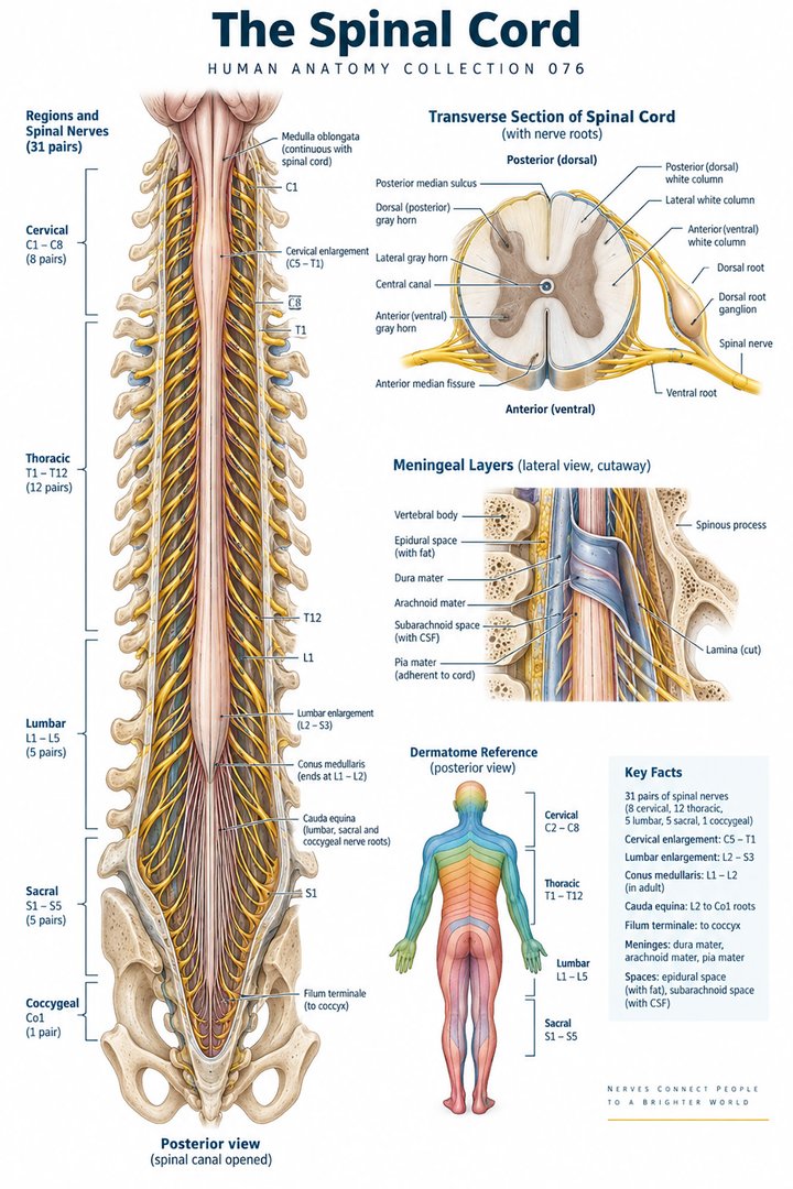 Human Spinal Cord ↗
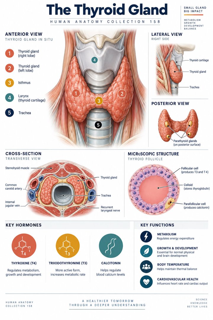 Human Thyroid Gland ↗
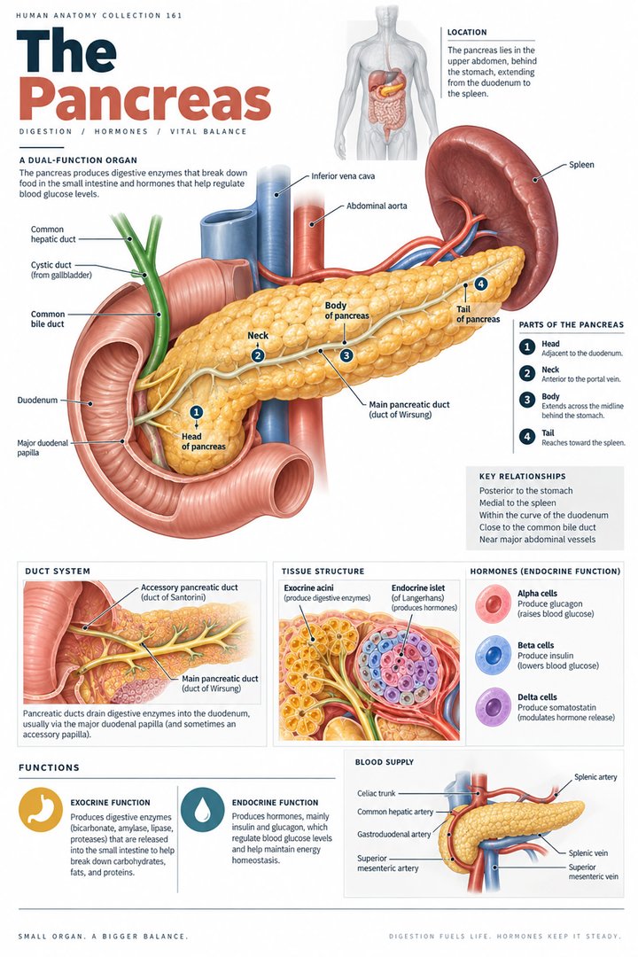 Human Pancreas Function ↗
 Human Gallbladder Anatomy ↗