The Human Eye
Individual poster · 300 DPI print files
Regular $9.90
Explore poster ↗$4.90USD
Offer price · Save $5.00Individual poster · 300 DPI print files
Reduced web preview. Separate high-resolution print files are supplied with the digital download.
ANATOMY 276
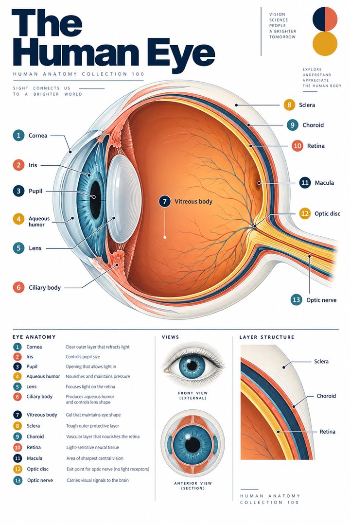
A modern anatomy illustration exploring the human eye, designed for educational and decorative use.
Purchases are not available yet. Checkout and download delivery are being prepared.
Sold by Kunstolymp.de · Germany
Compare prices & the complete collection ↗This is a DIGITAL DOWNLOAD. No physical product will be shipped. After purchase, download your files and print them at home, at a local print shop, or through an online professional printing service.
The separate 300 DPI print files are ideal for 24×36-inch posters (approximately 60×90 cm). ISO and 5×7 formats are included. The white border lets you adjust the layout or trim to fit other frame sizes; check the preview before printing to keep the illustration and labels visible.
The planned bundle delivery includes a PDF information sheet with a link to the relevant Google Drive folder and download instructions. The store is currently in preview and purchases are not yet available.
A science-inspired choice for a student’s study corner, an educator’s classroom, or an anatomy enthusiast’s wall. Final licensing terms will apply.
CLINICAL CONTEXT
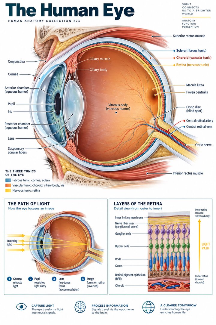
The eye converts light into neural signals and works with the brain to create vision. The cornea and lens focus incoming light; the iris regulates pupil size; the aqueous humour supports anterior-segment pressure and nutrition; the vitreous fills the posterior cavity; and the retina transduces light before signals pass through the optic nerve and visual pathways. Extraocular muscles align gaze, while eyelids and tear film protect the surface. A whole-eye diagram helps clinicians localise symptoms and connect structure with function. Blurred vision may arise from refractive error, corneal or lens disease, retinal pathology, optic neuropathy, or central neurological disease, so laterality, onset, pain, field change, and associated symptoms matter.
Refractive errors cause blurred distance or near vision and are often corrected with spectacles, contact lenses, or selected procedures. Dry eye and blepharitis may cause burning, grittiness, fluctuating vision, and tearing. Cataract gradually clouds the lens, reducing contrast and increasing glare. Glaucoma damages the optic nerve, often without early symptoms; chronic pressure-related injury differs from acute angle closure, which can cause severe eye pain, redness, halos, headache, nausea, and rapid visual loss. Age-related macular degeneration affects central vision and detail, while diabetic eye disease can injure retinal vessels. Conjunctivitis causes redness and discharge, but painful red eye with photophobia or reduced vision raises concern for keratitis, uveitis, or other urgent disease. Ask about trauma, contact lenses, diabetes, vascular risks, medicines, family history, and effect on reading, mobility, driving, and work.
Assess visual acuity in each eye, pupils, visual fields, ocular motility, external structures, and red reflex; use fluorescein, slit-lamp examination, tonometry, fundoscopy, or imaging when indicated and within competence. Sudden monocular loss, retinal symptoms, trauma, chemical exposure, or a painful red eye with reduced vision merits urgent ophthalmic evaluation. Treatment follows diagnosis: refractive correction and surface lubrication for selected symptoms; topical pressure-lowering medicines, laser, or surgery for glaucoma; cataract surgery when functional impairment warrants it; and anti-VEGF injections or laser for selected retinal disease. Diabetes and hypertension control, smoking cessation, protective eyewear, and recommended eye examinations help prevent or detect vision loss. Patients with diabetes need regular retinal screening even when vision feels normal.
Good lighting, screen breaks, hydration, and warm compresses for some eyelid conditions may support comfort but do not cure glaucoma, cataract, macular degeneration, or retinal disease. A nutrient-rich diet supports general health; AREDS2 supplements benefit particular stages of AMD and are not universal prevention. High-dose vitamins, herbal drops, or unregulated products can cause harm or interact with medicines; discuss them with an eye-care professional. Never put non-sterile substances in the eye or delay treatment for a “natural” remedy. Sudden vision loss, flashes with a shower of floaters, a curtain over vision, severe pain, chemical injury, or new double vision with neurological symptoms needs immediate assessment. This guide is educational and not an individual diagnosis.
Further reading: National Eye Institute: eye conditions · NEI: AREDS/AREDS2 supplements
Explain the working diagnosis, important alternatives, planned tests, expected course, and specific safety-netting in language the patient can use. Invite questions about treatment goals, capacity, cost, culture, and complementary therapies. Use a professional interpreter when language access could affect consent or understanding, and confirm clear arrangements for timely clinical review in writing. Document the reasoning, reassess response to treatment, and coordinate care across professions when illness affects more than one system. Teaching anatomy is most useful when it supports this partnership: accurate localisation, respectful listening, timely escalation, and a plan that remains responsive to the individual patient.
HUMAN ANATOMY COLLECTION · 001—300
A closer look at what makes us human. Discover anatomy posters for your study space, classroom, and everyday curiosity.
DIGITAL DOWNLOADS · 300 DPI PRINT FILES
EXPLORE BY SYSTEM
LEARN & EXPLORE
Detailed English guides with Latin terminology, diagrams, real images, and clinical learning connections.
 Human Anatomy Diagrams ↗
 Human Body Organs ↗
 Skeletal System Anatomy ↗
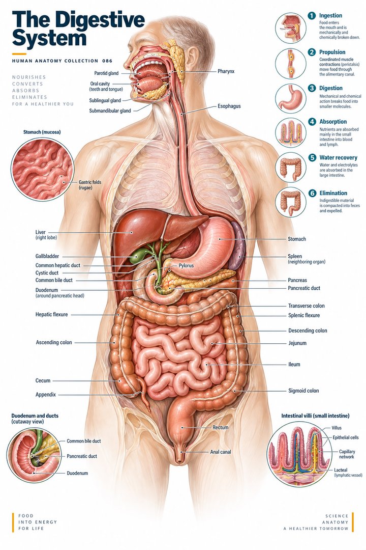 Human Digestive System ↗
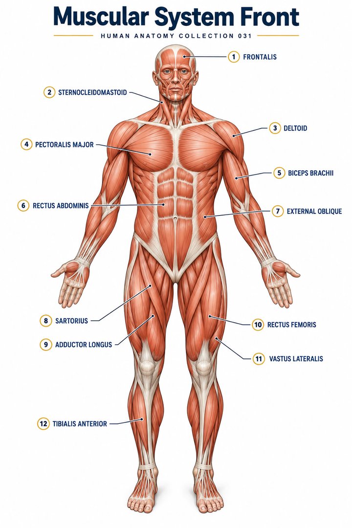 Muscular System Overview ↗
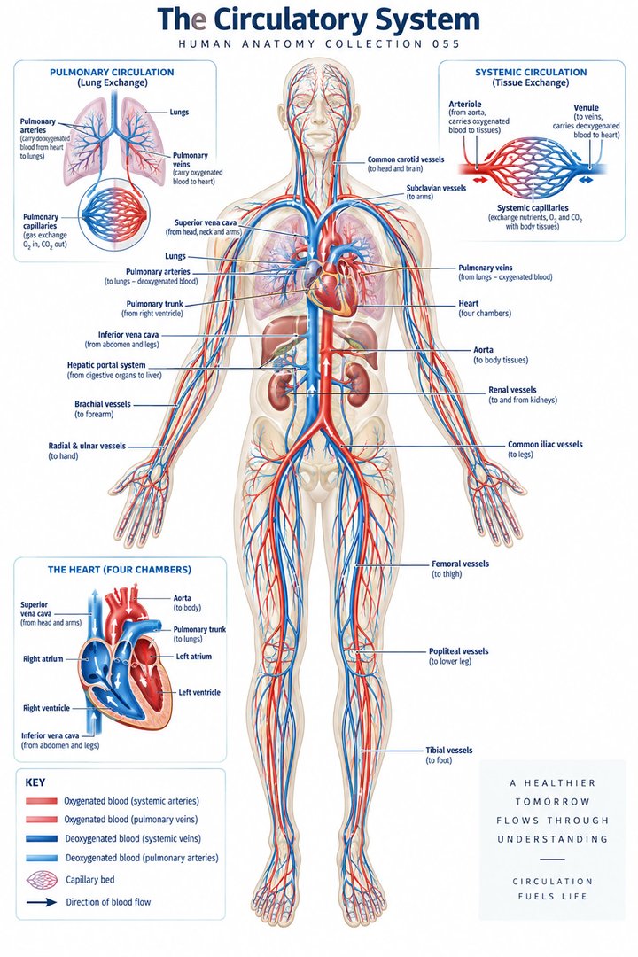 Human Circulatory System ↗
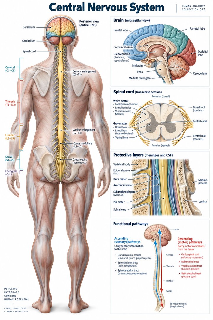 Human Nervous System ↗
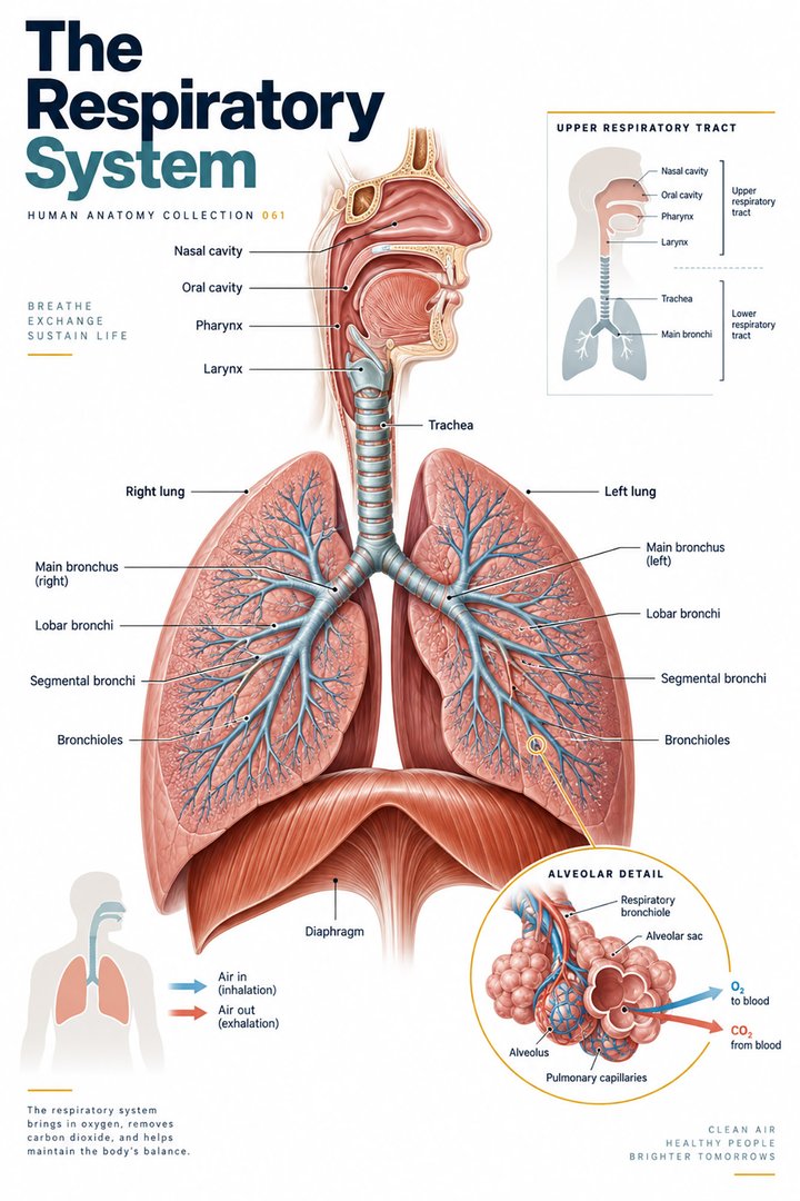 Respiratory System Functions ↗
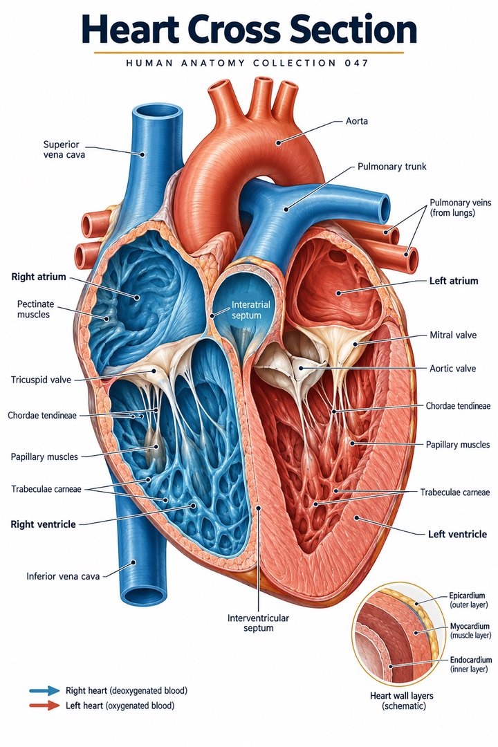 Human Heart Structure ↗
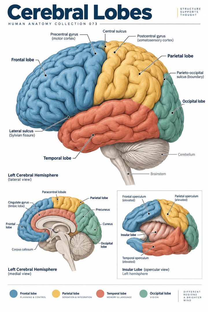 Human Brain Lobes ↗
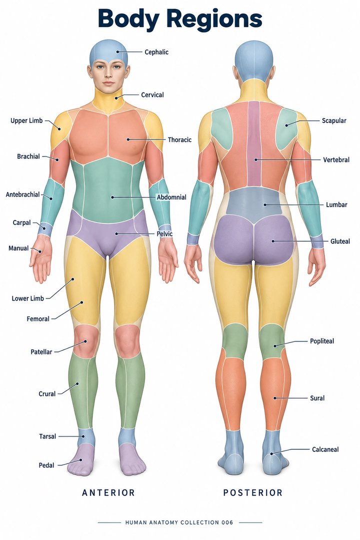 Human Body Parts ↗
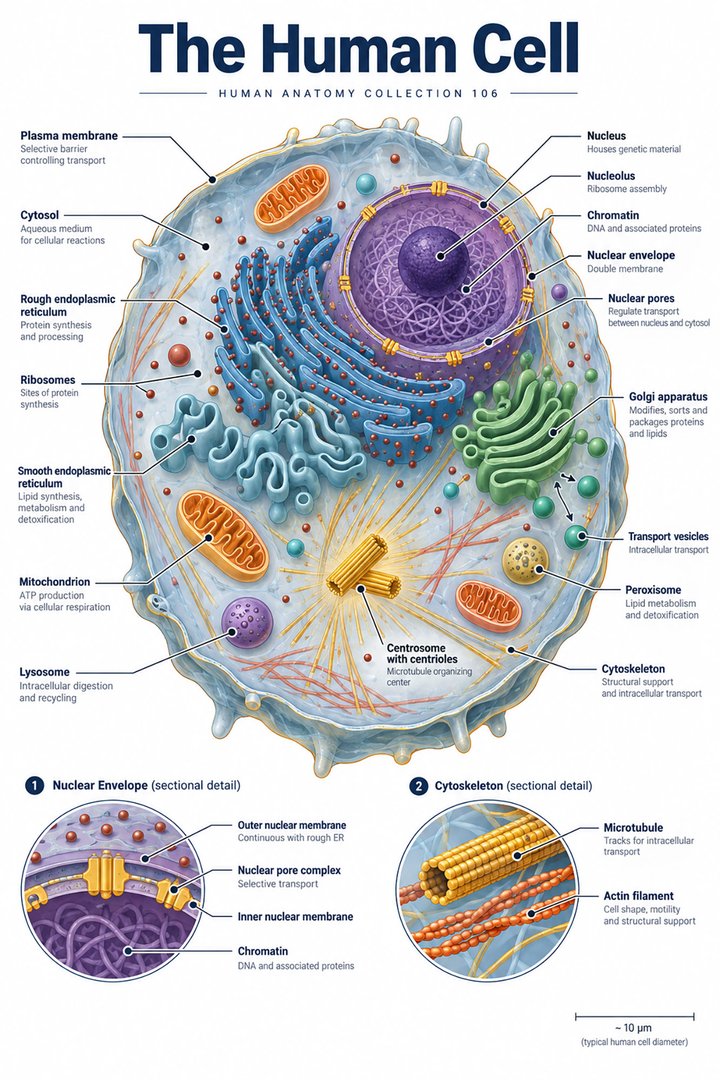 Human Cell Structure ↗
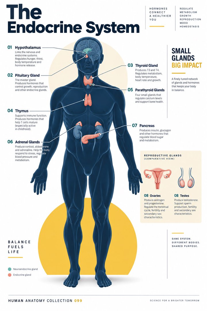 Human Endocrine System ↗
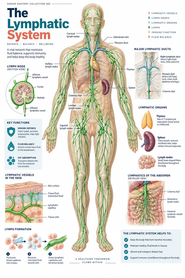 Human Lymphatic System ↗
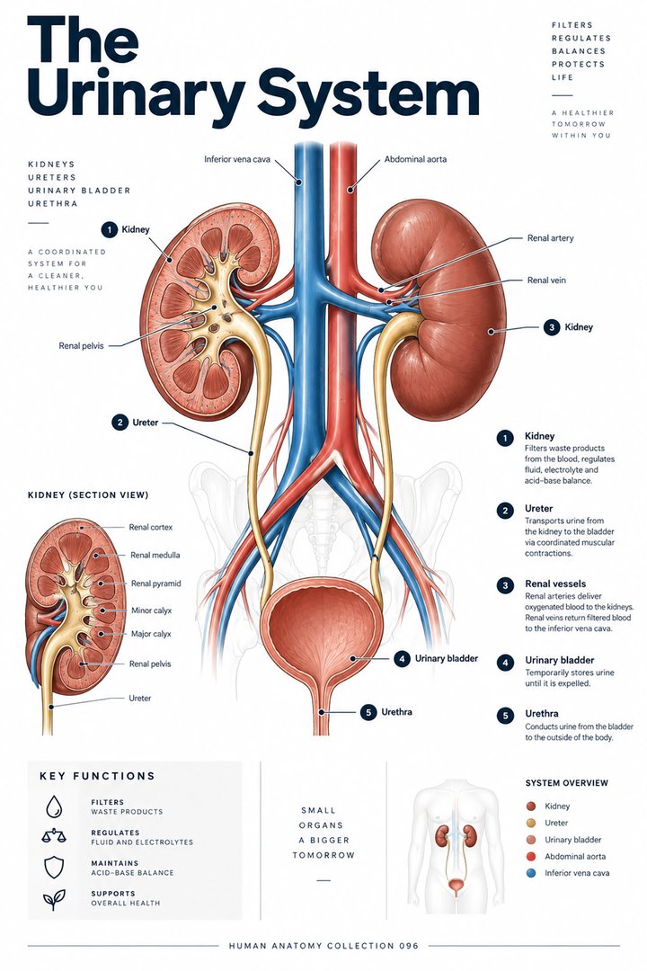 Human Urinary System ↗
 Male Reproductive System ↗
 Female Reproductive System ↗
 Human Immune System ↗Central Nervous System ↗
 Human Eye Structure ↗
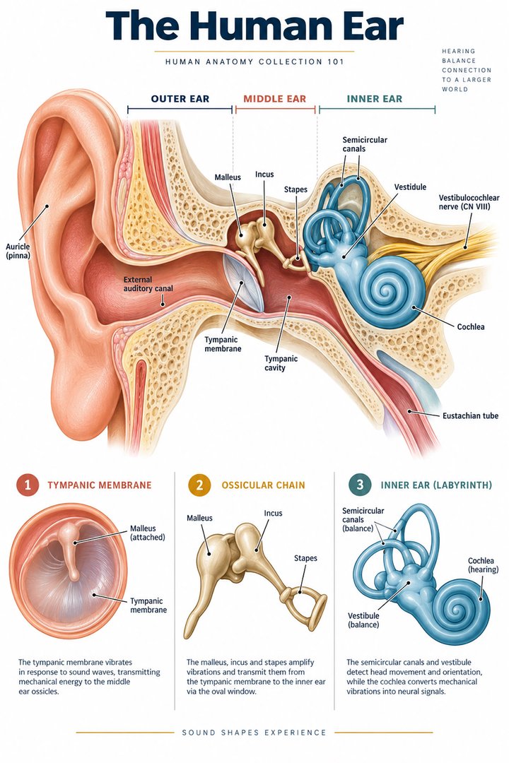 Human Ear Structure ↗
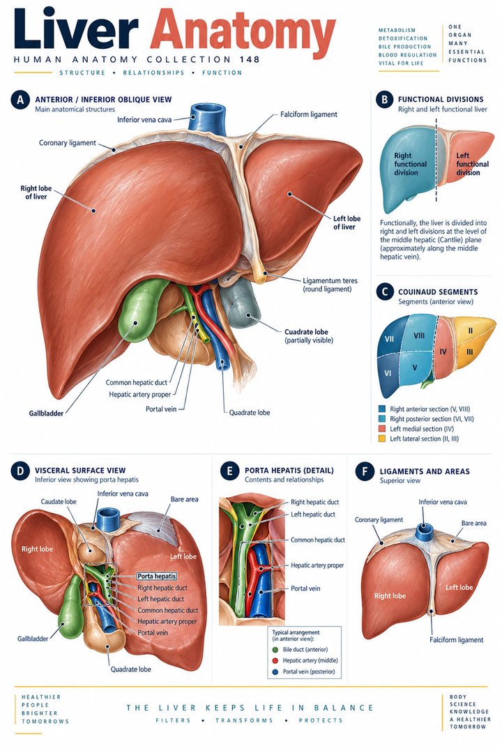 Human Liver Function ↗
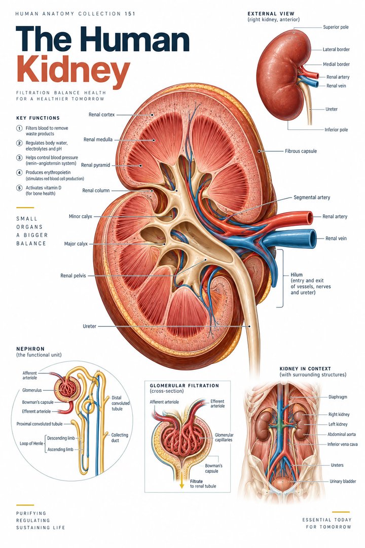 Human Kidney Anatomy ↗
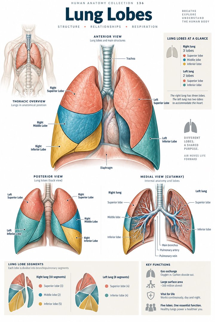 Human Lung Anatomy ↗
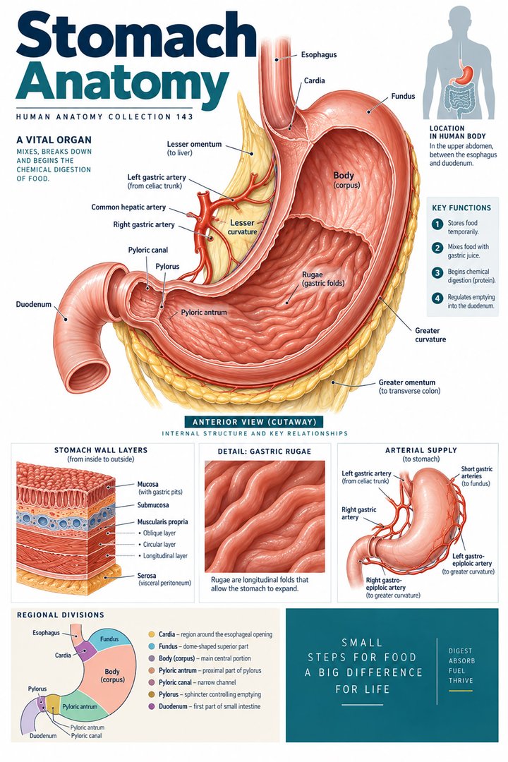 Human Stomach Structure ↗
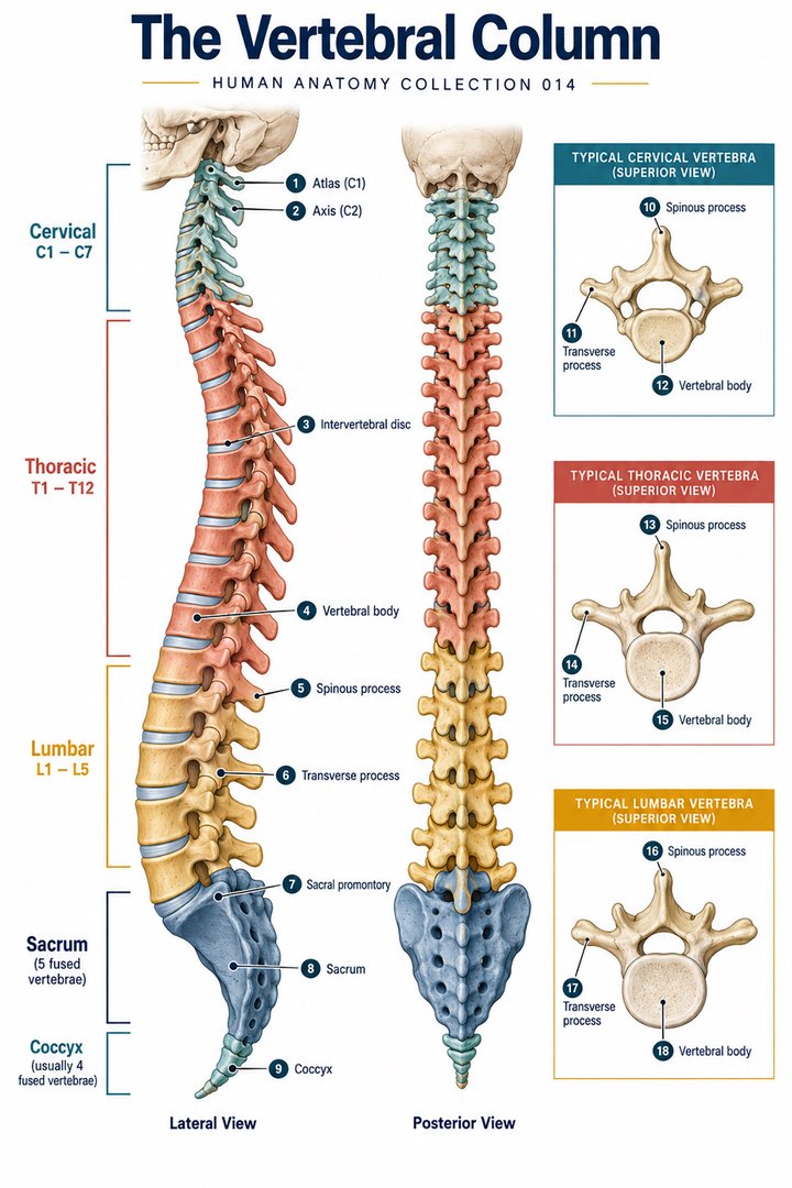 Human Spine Anatomy ↗
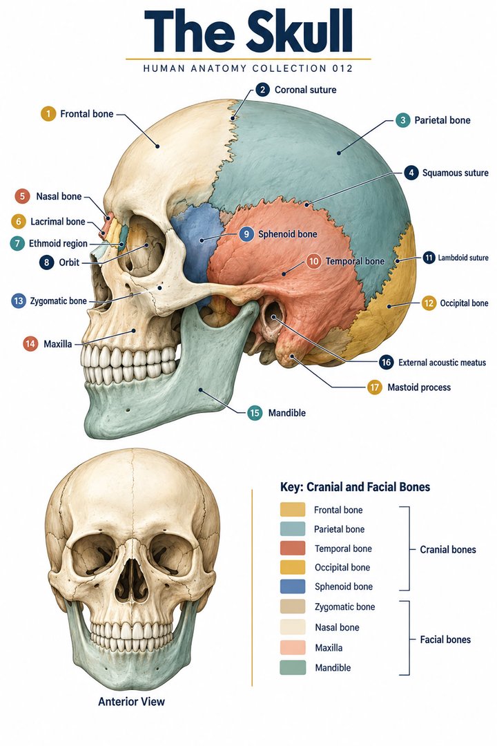 Human Skull Anatomy ↗
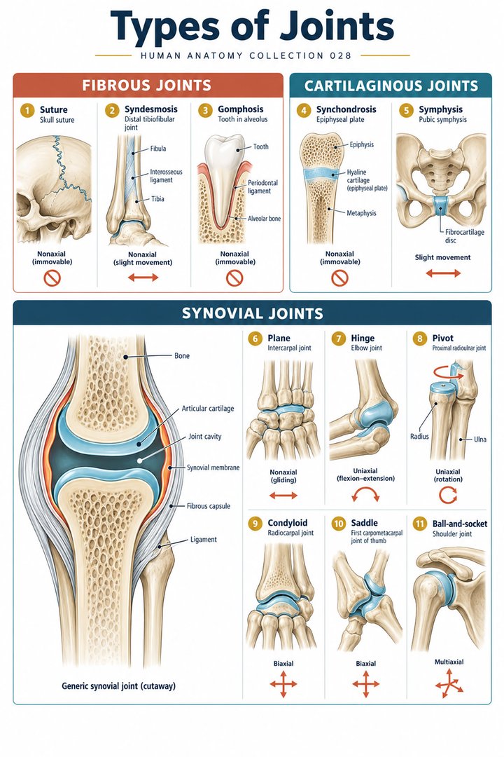 Human Joint Types ↗
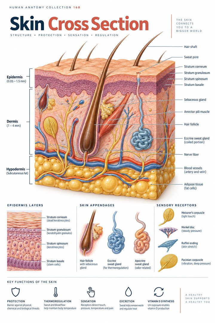 Human Skin Layers ↗
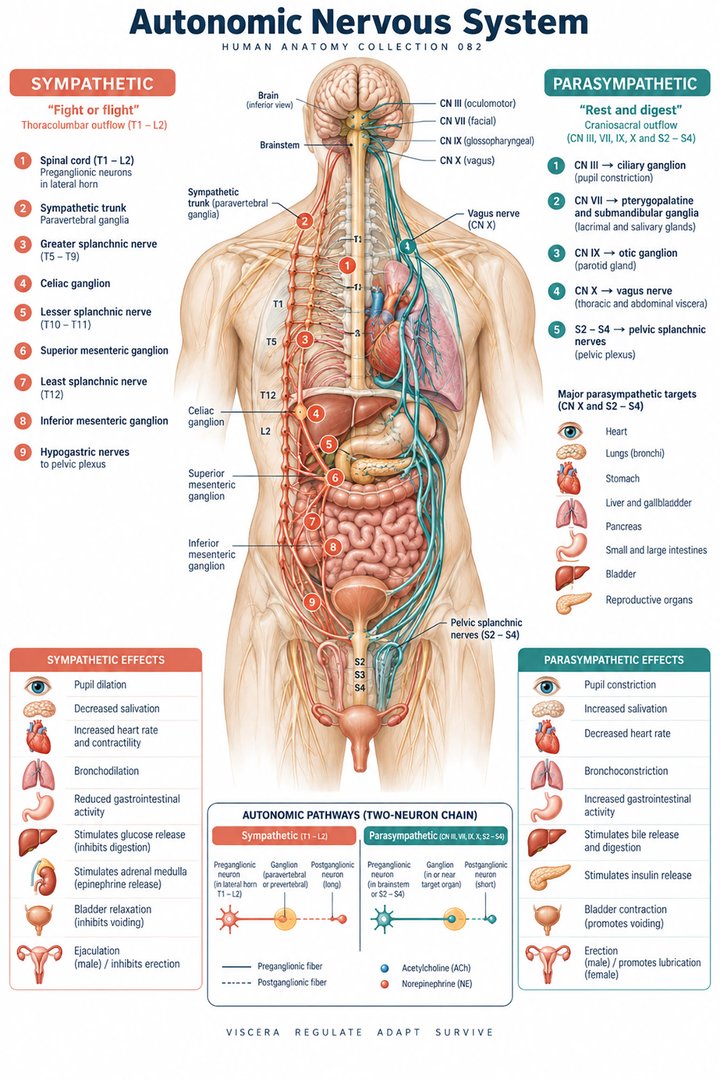 Autonomic Nervous System ↗
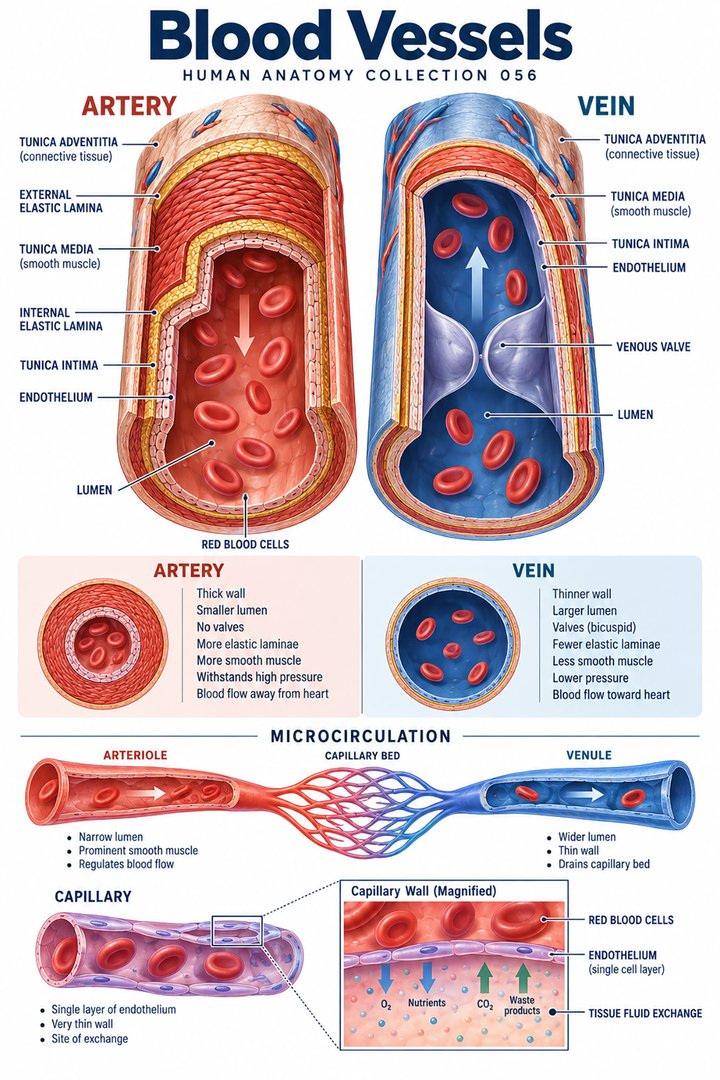 Human Blood Vessels ↗
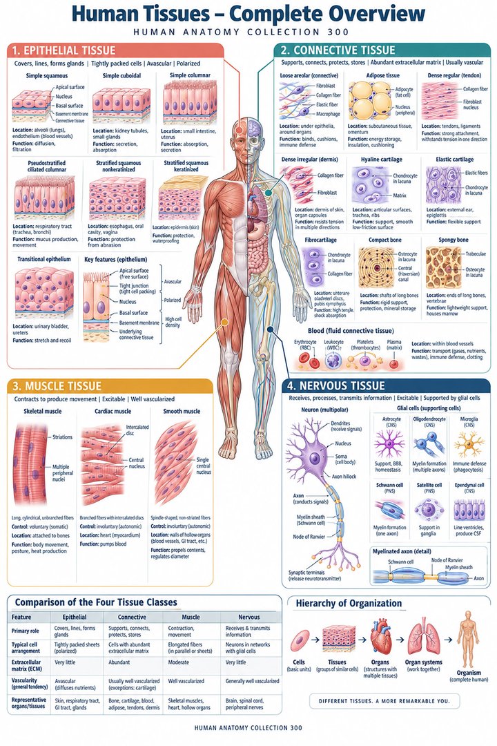 Human Body Tissues ↗
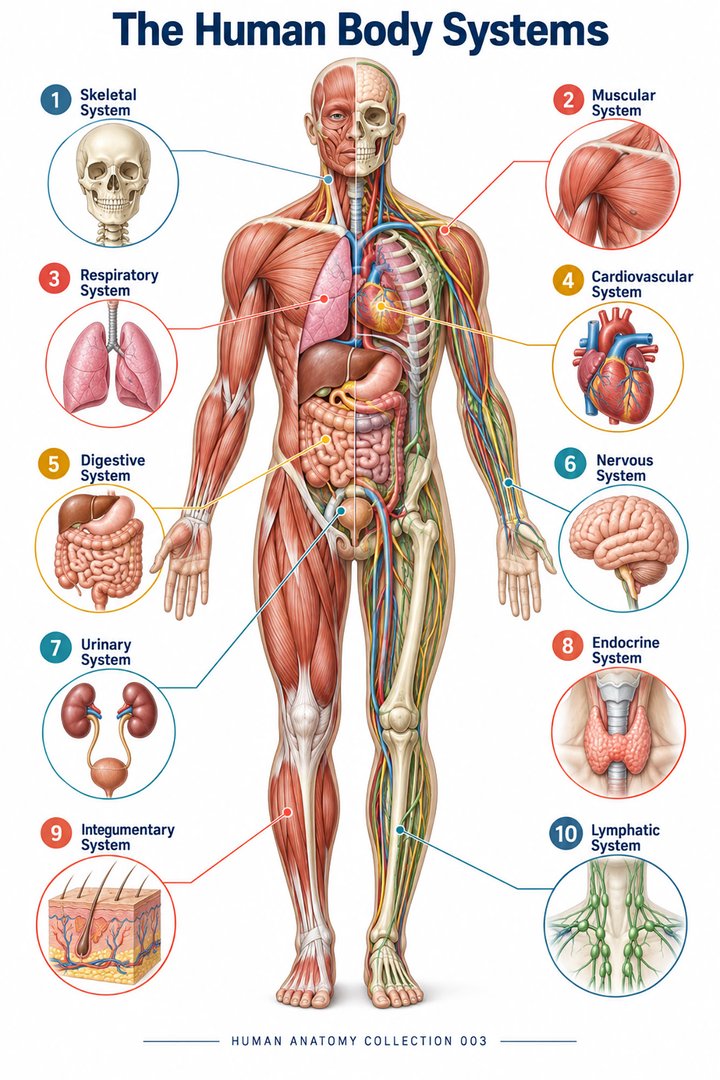 Human Body Systems ↗
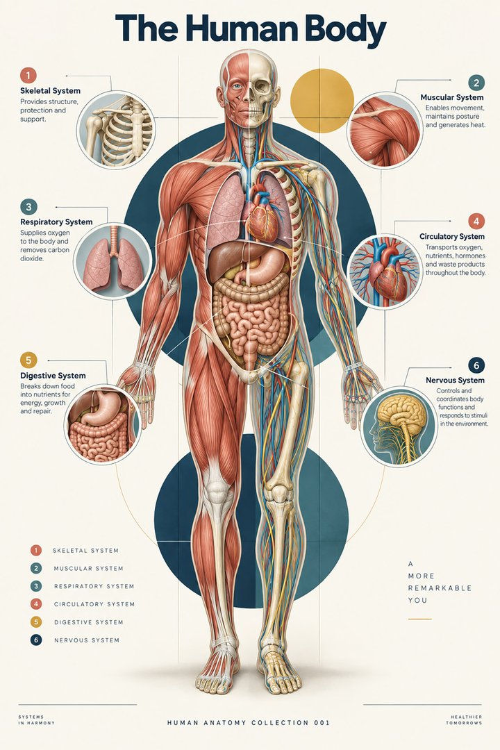 Human Body Facts ↗Human Anatomy and Physiology ↗
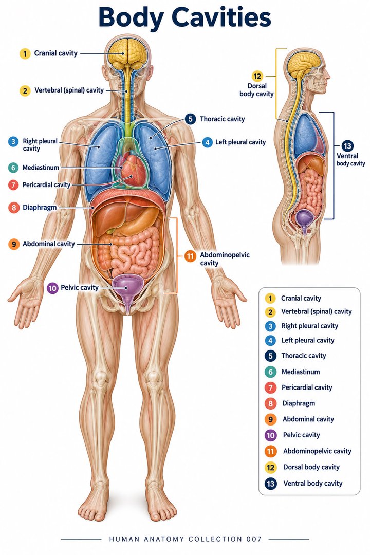 Human Body Cavities ↗Human Torso Anatomy ↗Human Digestive Tract ↗
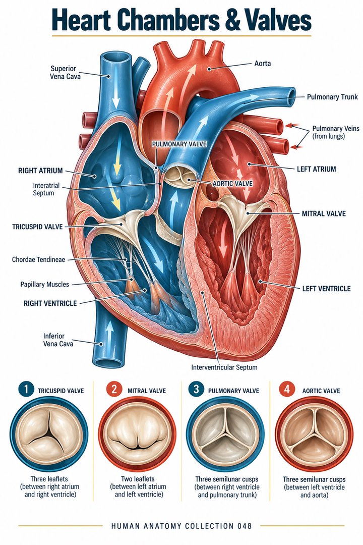 Human Heart Chambers ↗Human Organ Placement ↗
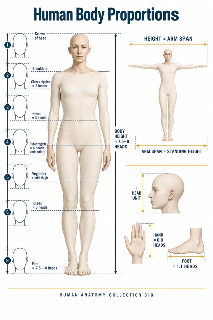 Human Body Proportions ↗
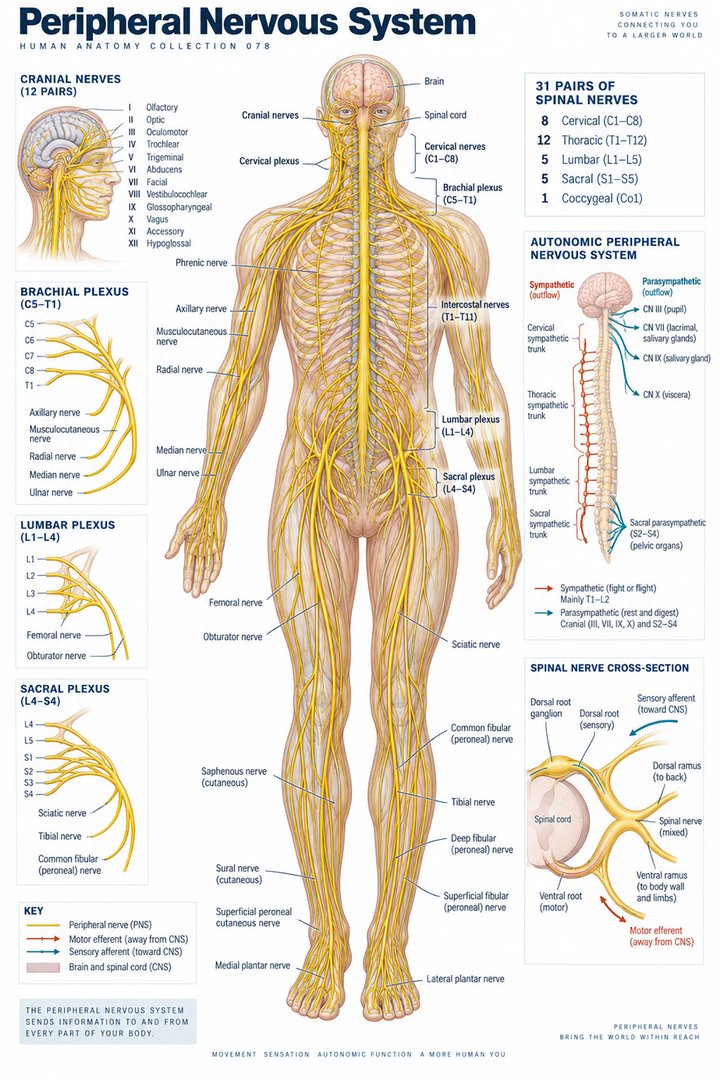 Peripheral Nervous System ↗
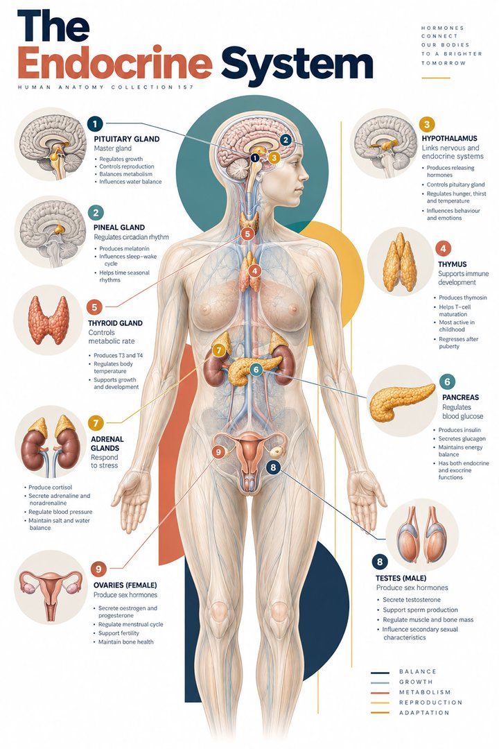 Human Endocrine Glands ↗
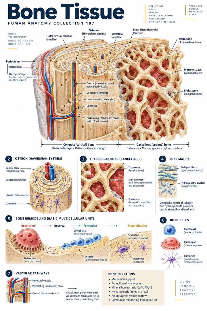 Human Bone Density ↗Human Blood Circulation ↗
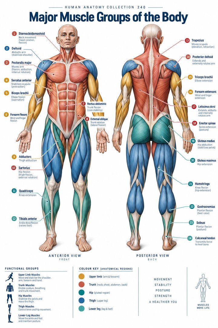 Human Muscle Groups ↗
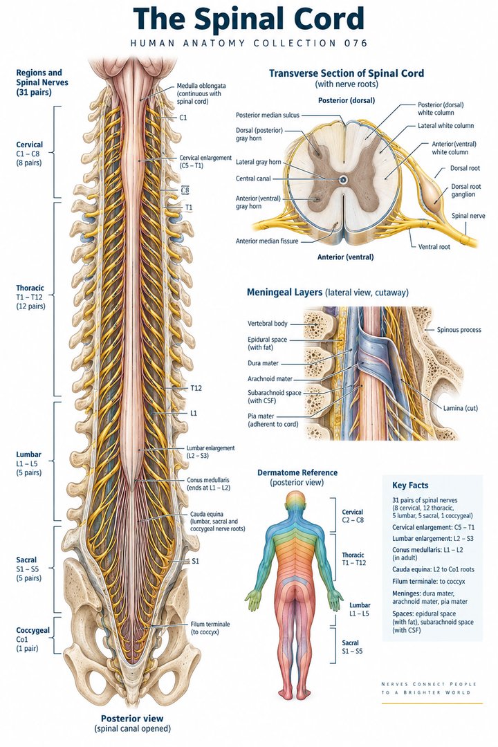 Human Spinal Cord ↗
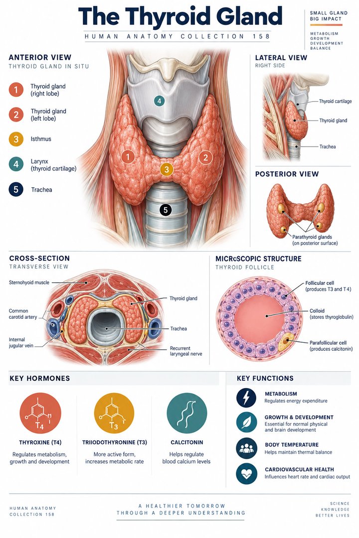 Human Thyroid Gland ↗
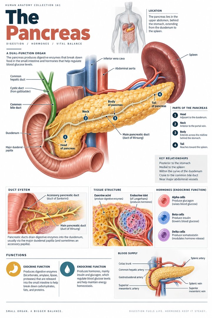 Human Pancreas Function ↗
 Human Gallbladder Anatomy ↗