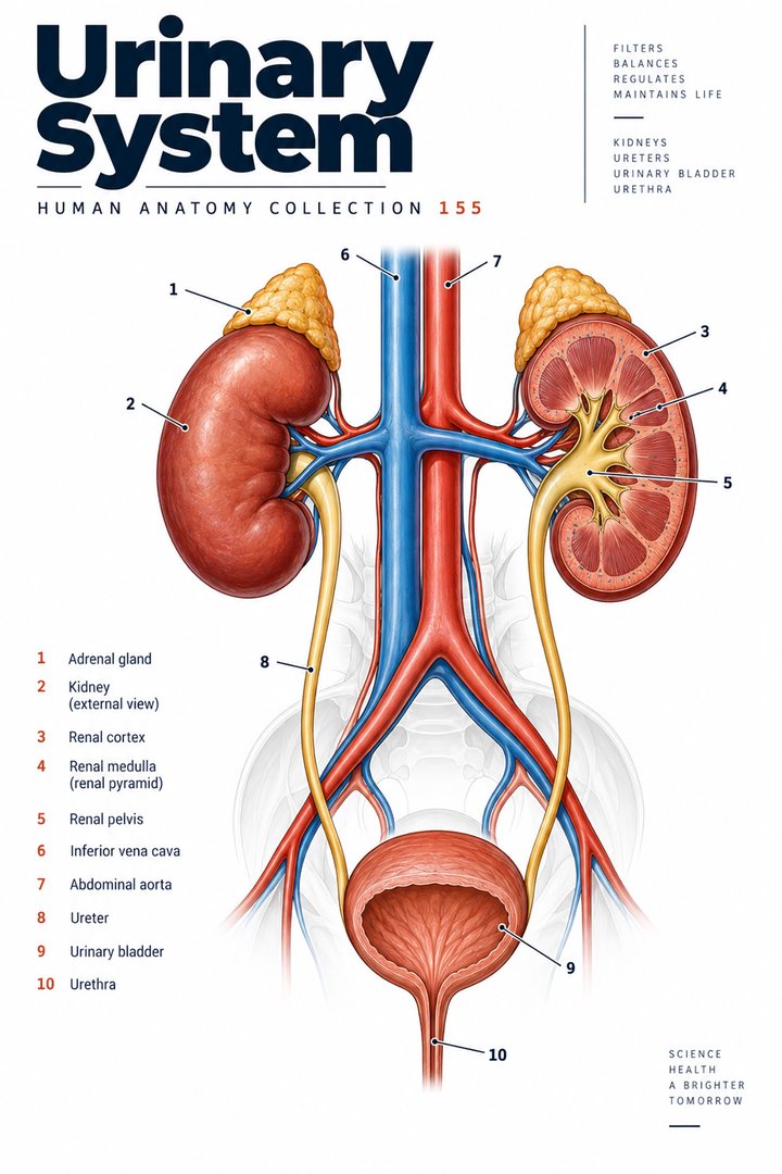
Urinary System
Individual poster · 300 DPI print files
Regular $9.90
Explore poster ↗$4.90USD
Offer price · Save $5.00Individual poster · 300 DPI print files
Reduced web preview. Separate high-resolution print files are supplied with the digital download.
ANATOMY 155
A modern anatomy illustration exploring urinary system, designed for educational and decorative use.
Purchases are not available yet. Checkout and download delivery are being prepared.
Sold by Kunstolymp.de · Germany
Compare prices & the complete collection ↗This is a DIGITAL DOWNLOAD. No physical product will be shipped. After purchase, download your files and print them at home, at a local print shop, or through an online professional printing service.
The separate 300 DPI print files are ideal for 24×36-inch posters (approximately 60×90 cm). ISO and 5×7 formats are included. The white border lets you adjust the layout or trim to fit other frame sizes; check the preview before printing to keep the illustration and labels visible.
The planned bundle delivery includes a PDF information sheet with a link to the relevant Google Drive folder and download instructions. The store is currently in preview and purchases are not yet available.
A science-inspired choice for a student’s study corner, an educator’s classroom, or an anatomy enthusiast’s wall. Final licensing terms will apply.
CLINICAL CONTEXT
The urinary system comprises kidneys, ureters, bladder, and urethra. Nephrons filter plasma, reclaim water and solutes, excrete waste, and help regulate electrolytes, acid-base balance, blood pressure, and erythropoietin. Urine passes through the collecting system and ureters to the bladder for storage and is released through coordinated detrusor contraction and sphincter relaxation. Anatomy helps localise symptoms: renal or ureteric disease may cause flank pain or haematuria; bladder inflammation may cause dysuria, urgency, and frequency; outlet obstruction can impair emptying. These patterns are not diagnostic by themselves, and reproductive, gastrointestinal, vascular, medication-related, and musculoskeletal causes may overlap.
Cystitis commonly causes painful, frequent urination and suprapubic discomfort. Fever, rigors, flank pain, nausea, or systemic illness raises concern for pyelonephritis, which may progress to sepsis, especially in pregnancy, older adults, or people with diabetes or impaired immunity. Urolithiasis can cause severe colicky flank pain radiating toward the groin, nausea, and blood in urine; obstruction with infection is an emergency. Chronic kidney disease may remain silent until advanced, so diabetes, hypertension, family history, albuminuria, and medication exposure matter. Lower urinary tract symptoms such as weak stream, hesitancy, nocturia, retention, or incontinence may reflect benign prostatic enlargement, pelvic floor dysfunction, neurological disease, infection, or medication effects. Ask about pregnancy, sexual and urinary history as appropriate, fluid intake, stone history, procedures, renal-toxic medicines, and effects on sleep and daily life.
Assessment includes vital signs, hydration, abdominal and flank examination, urinalysis and culture when indicated, renal function, electrolytes, urine albumin, pregnancy testing where relevant, and imaging for suspected obstruction, stone, recurrent infection, or structural disease. Treatment is cause-specific: antibiotics for supported bacterial infection, analgesia and selected expulsive or procedural management for stones, and risk-factor and medication management for CKD. Obstructed infected systems need urgent drainage and antimicrobial care. CKD management may include blood-pressure and diabetes control, kidney-protective medicines, dietary review, vaccination, and nephrology referral based on severity. Retention may require bladder scanning, catheterisation, and evaluation of the underlying cause. Avoid assuming all urinary symptoms represent infection or prescribing from symptoms alone when testing or examination is needed.
Appropriate hydration, smoking cessation, movement, and individualised nutrition can support general and kidney health; fluid targets must account for heart failure, kidney failure, and clinician advice. Cranberry products may modestly reduce recurrent UTI risk for some people but do not treat an active kidney infection. Herbal remedies and “kidney cleanses” may contain nephrotoxic ingredients or interact with medicines. Discuss supplements openly and avoid stopping prescribed treatment. Urgent care is needed for fever with flank pain, vomiting with inability to hydrate, visible blood with clots, inability to urinate, severe colic with fever, confusion, fainting, or rapidly worsening illness. This guide is educational, not individual medical advice.
Further reading: NIDDK: kidney infection · NIDDK: kidney stones · NIDDK: chronic kidney disease
Explain the working diagnosis, important alternatives, planned tests, expected course, and specific safety-netting in language the patient can use. Invite questions about treatment goals, capacity, cost, culture, and complementary therapies. Use a professional interpreter when language access could affect consent or understanding, and confirm clear arrangements for timely clinical review in writing. Document the reasoning, reassess response to treatment, and coordinate care across professions when illness affects more than one system. Teaching anatomy is most useful when it supports this partnership: accurate localisation, respectful listening, timely escalation, and a plan that remains responsive to the individual patient.
HUMAN ANATOMY COLLECTION · 001—300
A closer look at what makes us human. Discover anatomy posters for your study space, classroom, and everyday curiosity.
DIGITAL DOWNLOADS · 300 DPI PRINT FILES
EXPLORE BY SYSTEM
LEARN & EXPLORE
Detailed English guides with Latin terminology, diagrams, real images, and clinical learning connections.
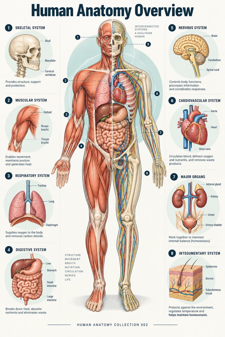 Human Anatomy Diagrams ↗
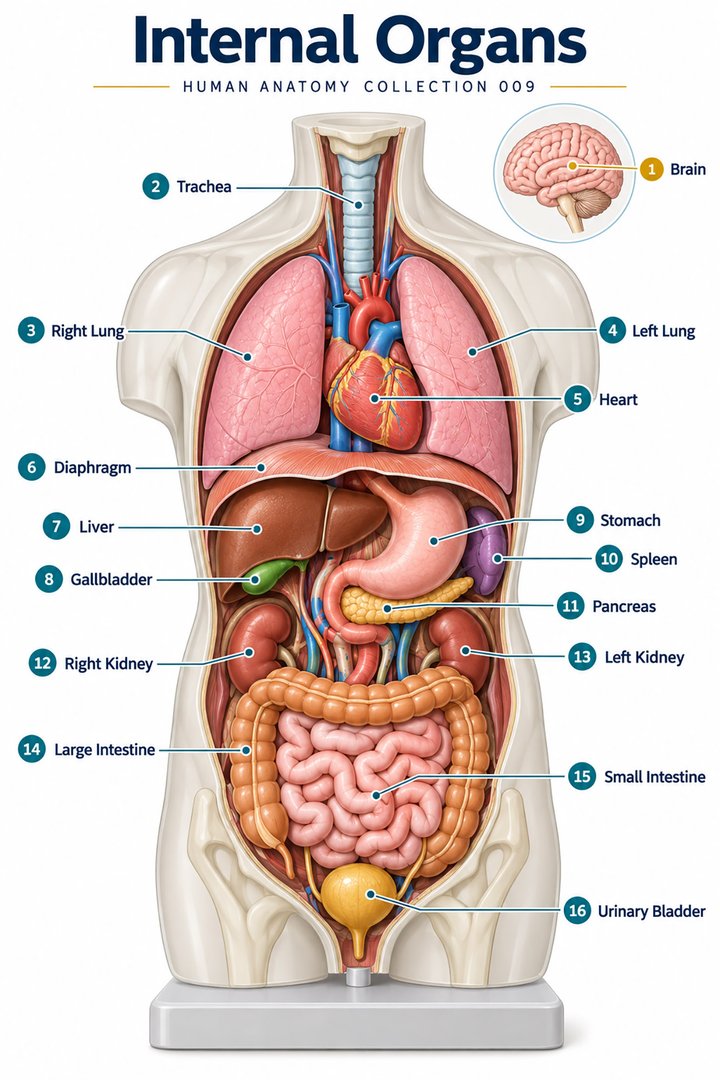 Human Body Organs ↗
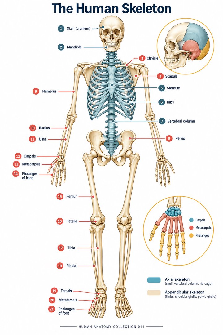 Skeletal System Anatomy ↗
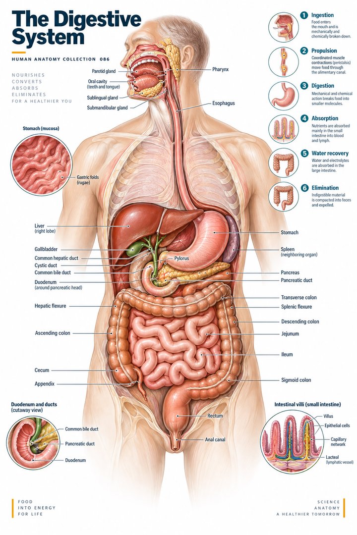 Human Digestive System ↗
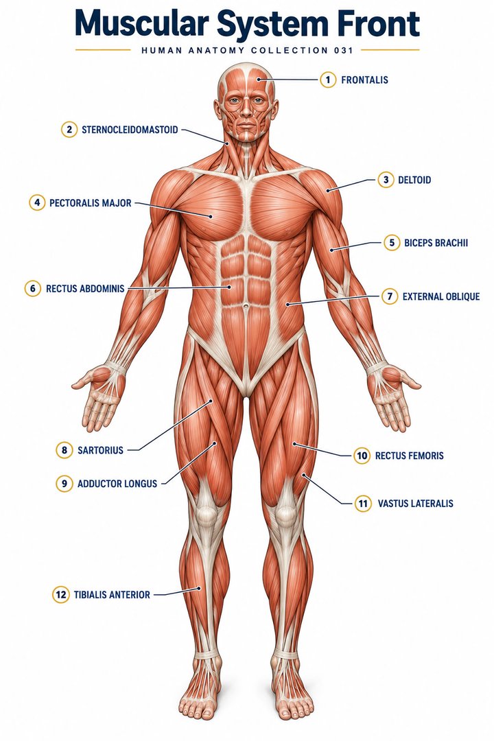 Muscular System Overview ↗
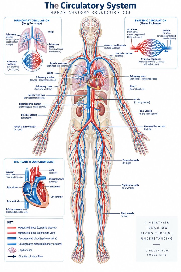 Human Circulatory System ↗
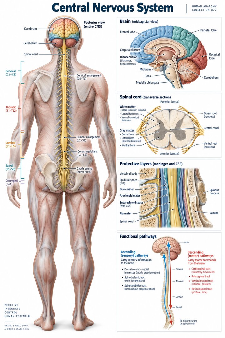 Human Nervous System ↗
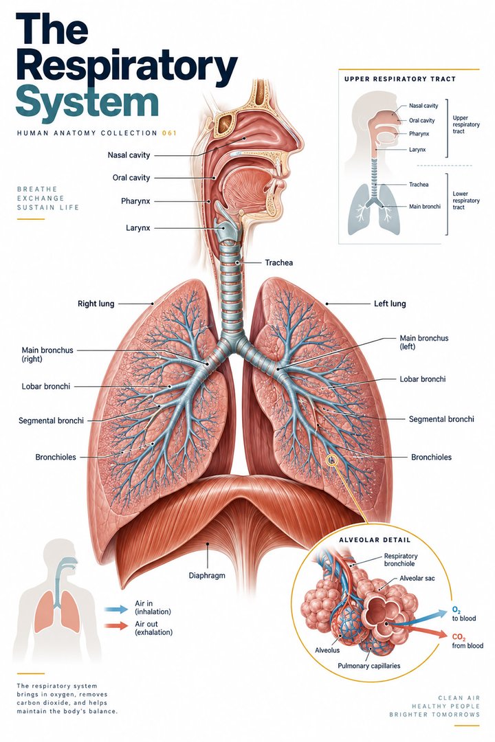 Respiratory System Functions ↗
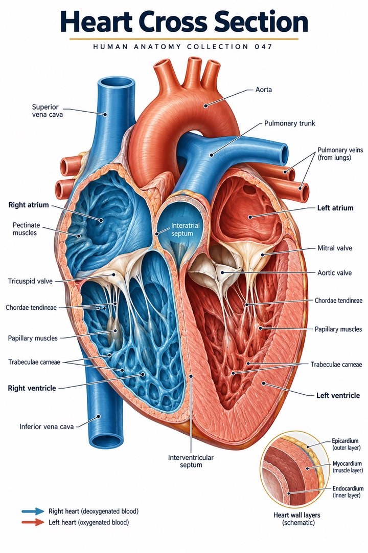 Human Heart Structure ↗
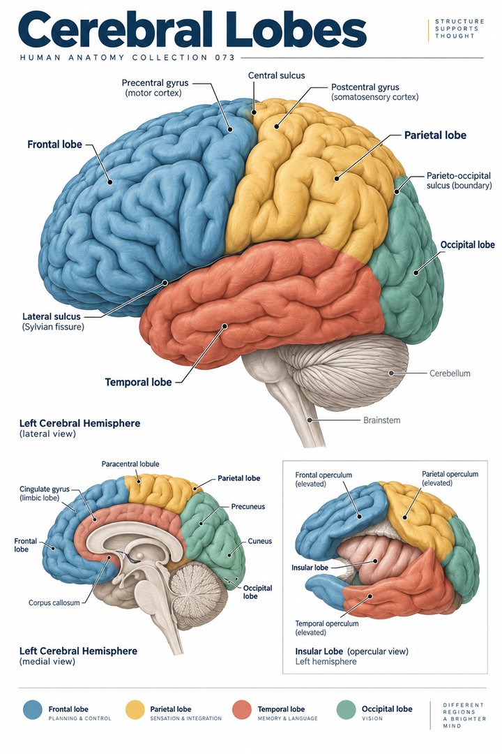 Human Brain Lobes ↗
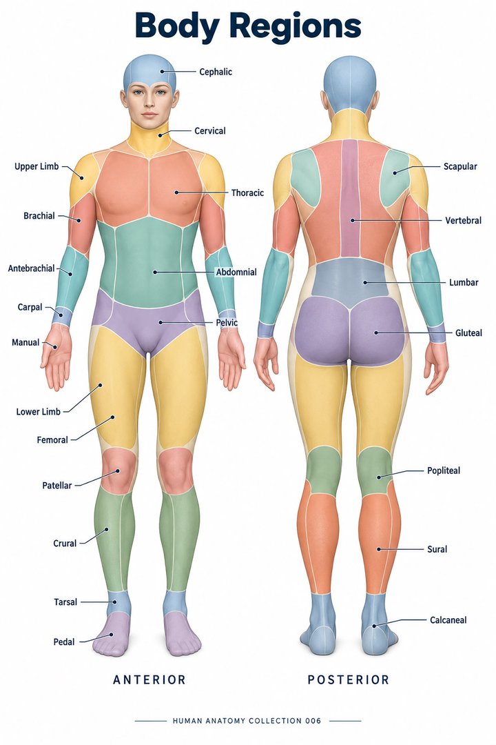 Human Body Parts ↗
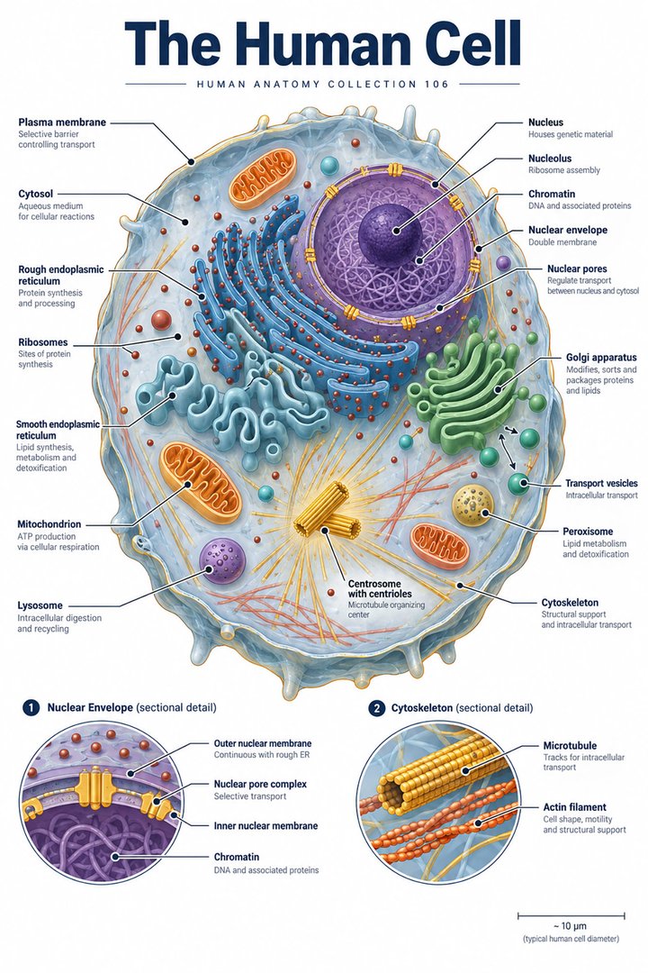 Human Cell Structure ↗
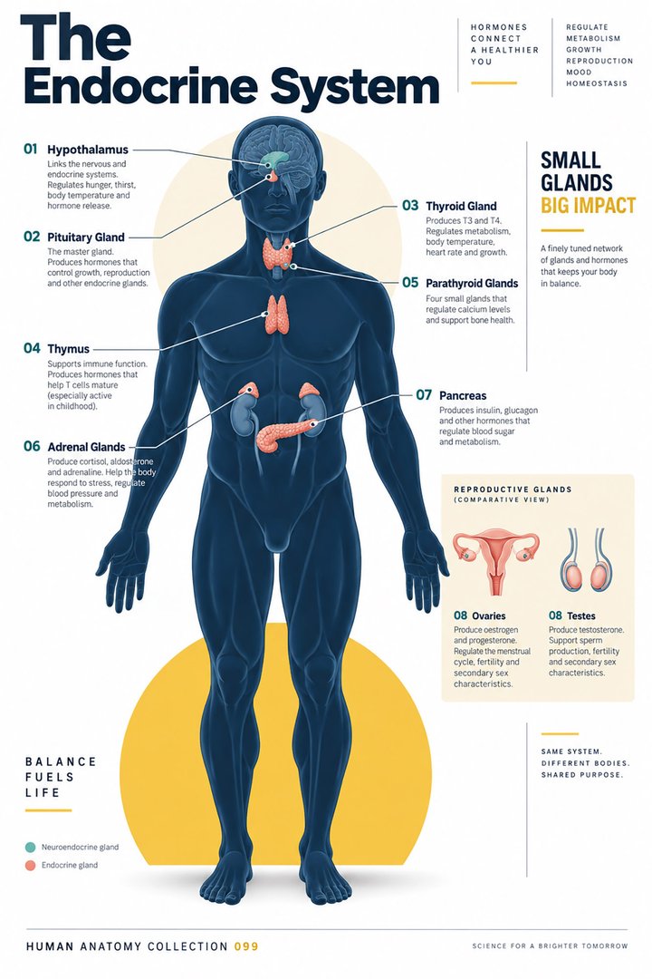 Human Endocrine System ↗
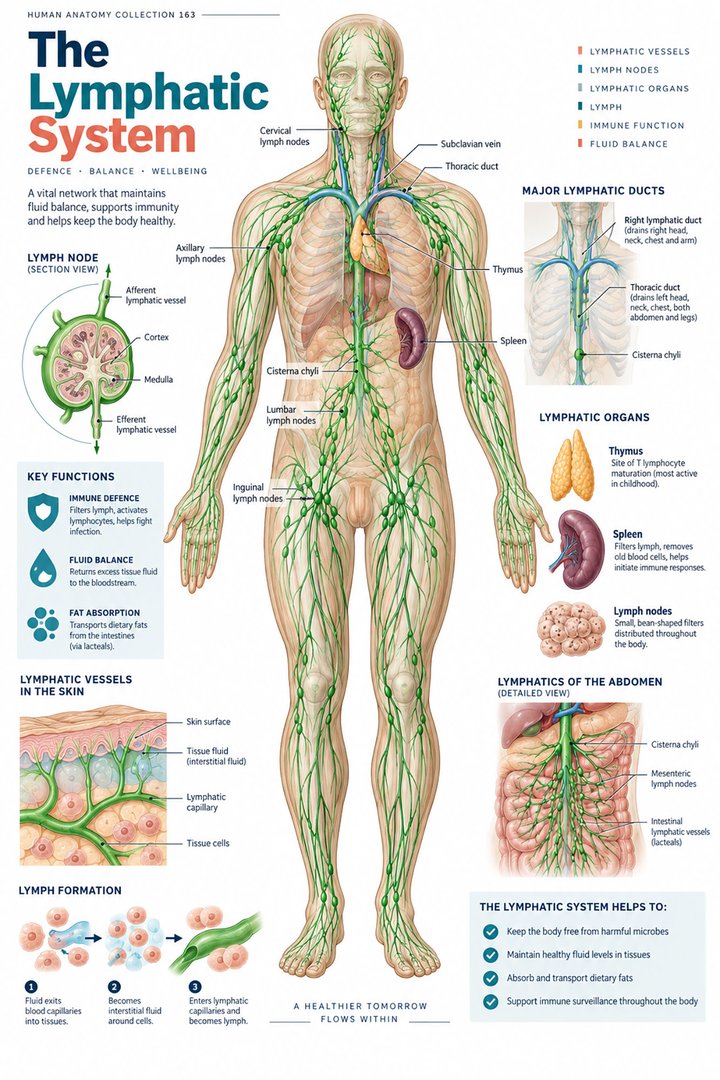 Human Lymphatic System ↗
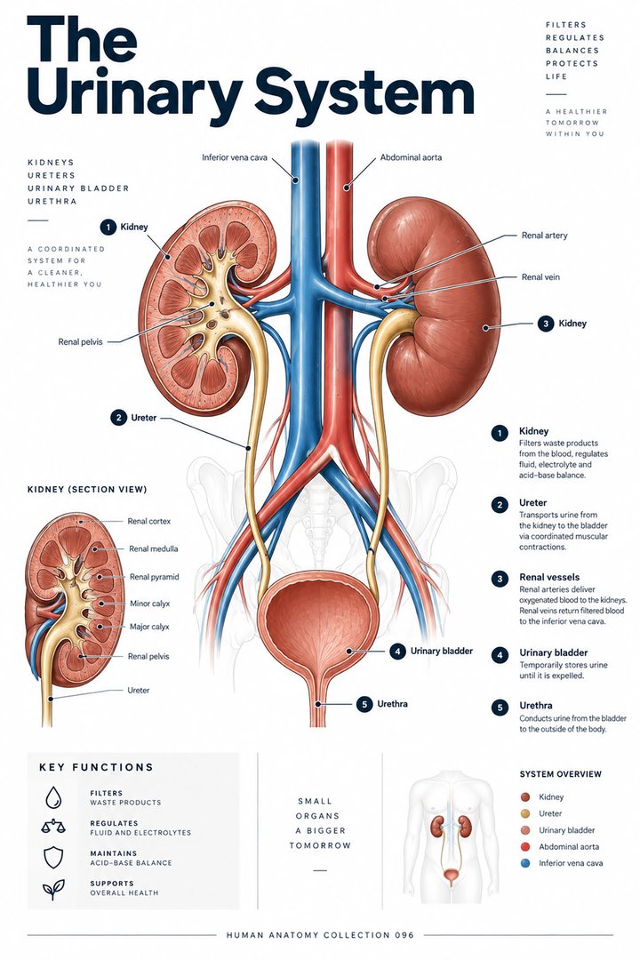 Human Urinary System ↗
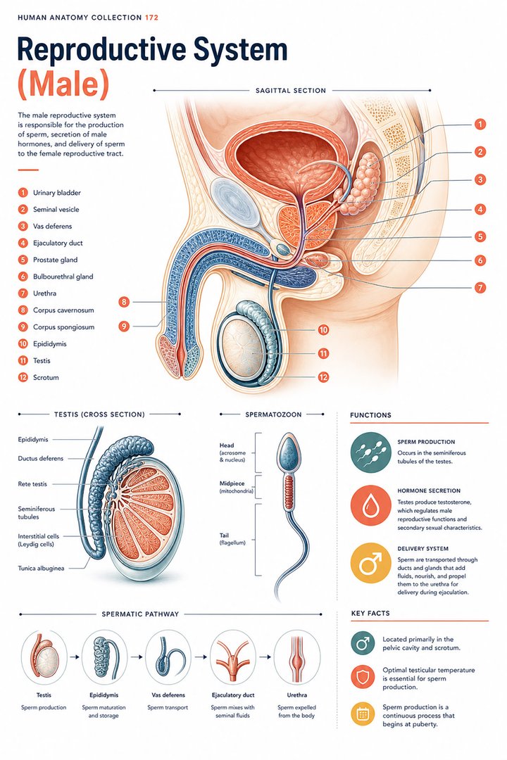 Male Reproductive System ↗
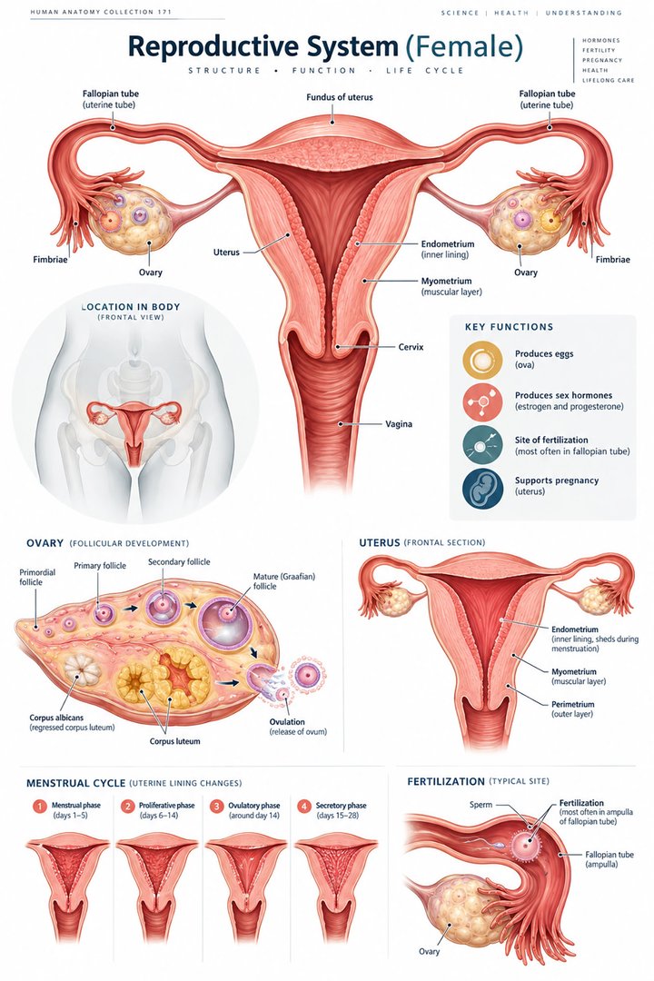 Female Reproductive System ↗
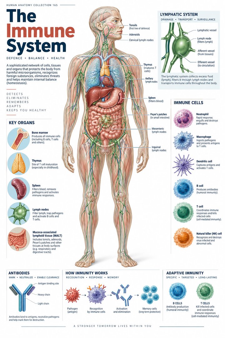 Human Immune System ↗Central Nervous System ↗
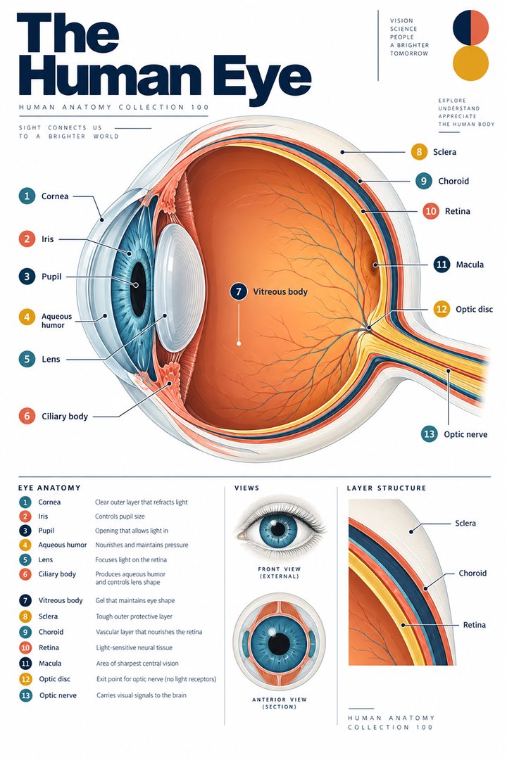 Human Eye Structure ↗
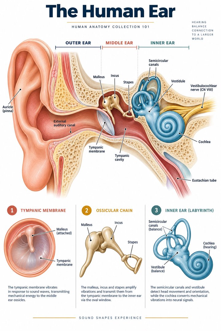 Human Ear Structure ↗
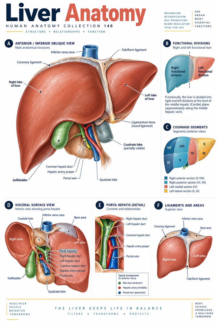 Human Liver Function ↗
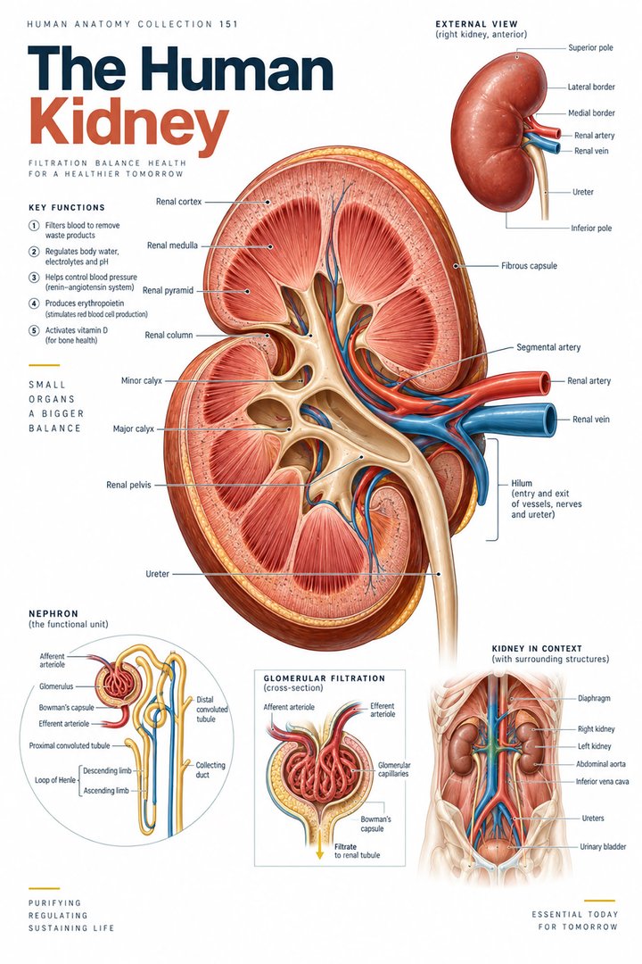 Human Kidney Anatomy ↗
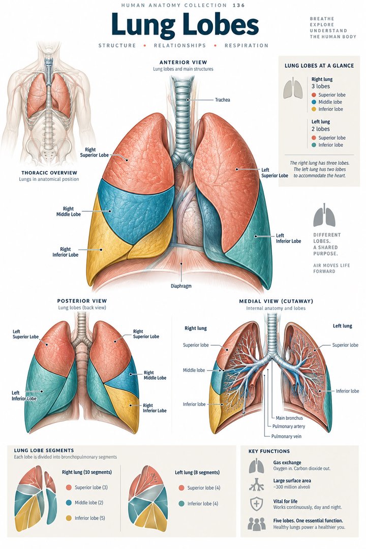 Human Lung Anatomy ↗
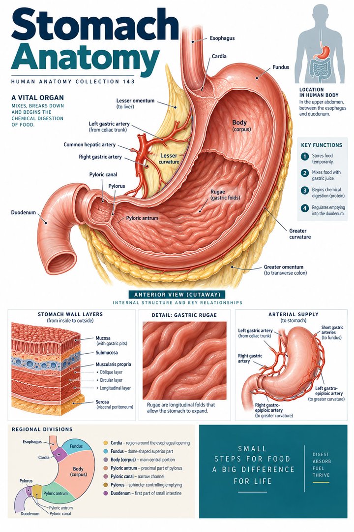 Human Stomach Structure ↗
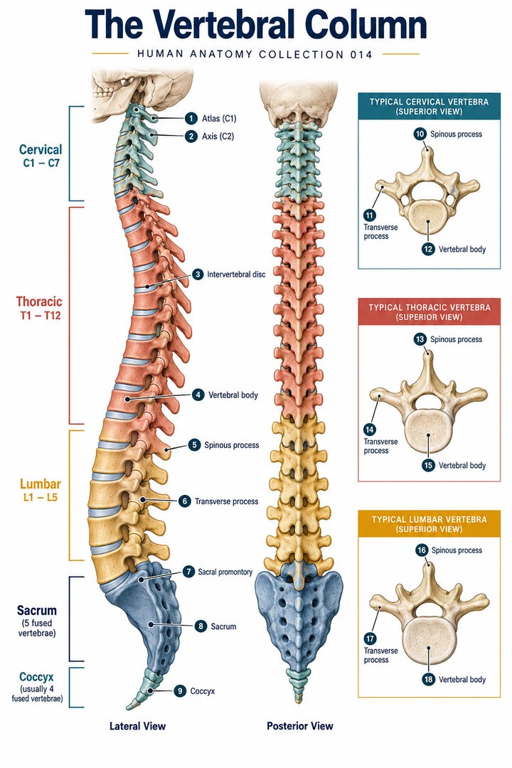 Human Spine Anatomy ↗
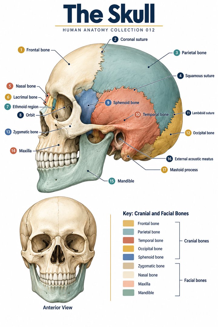 Human Skull Anatomy ↗
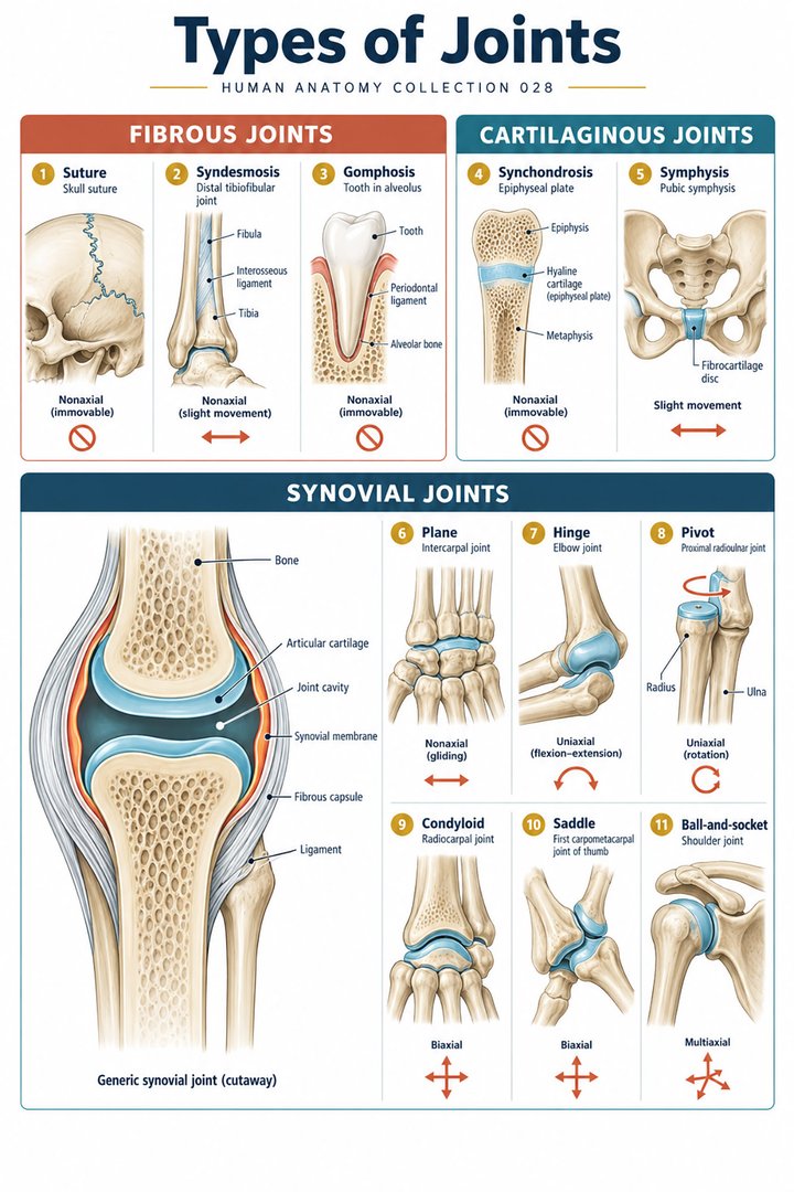 Human Joint Types ↗
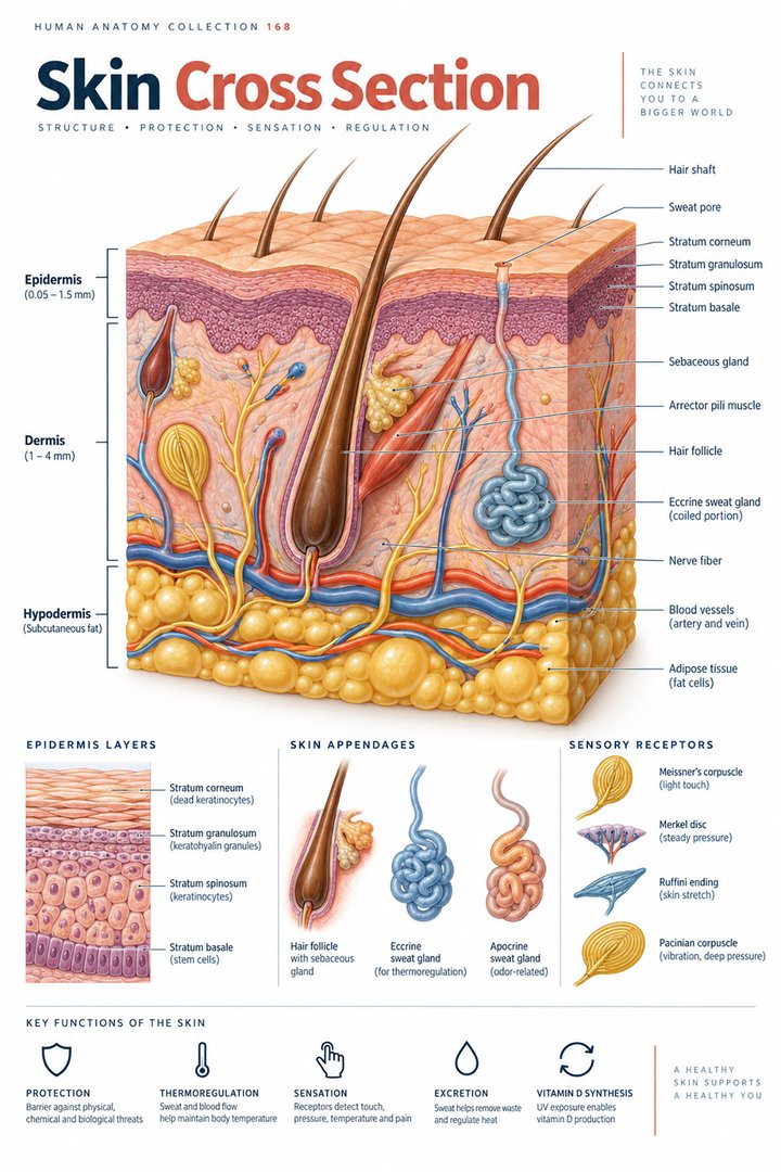 Human Skin Layers ↗
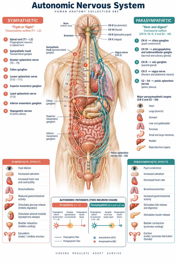 Autonomic Nervous System ↗
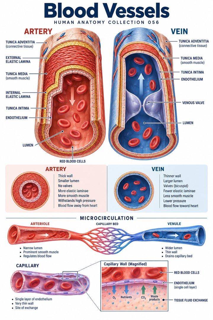 Human Blood Vessels ↗
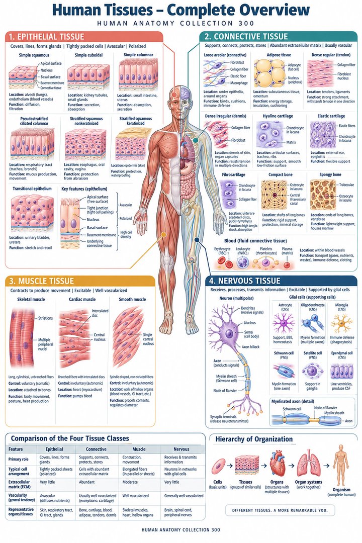 Human Body Tissues ↗
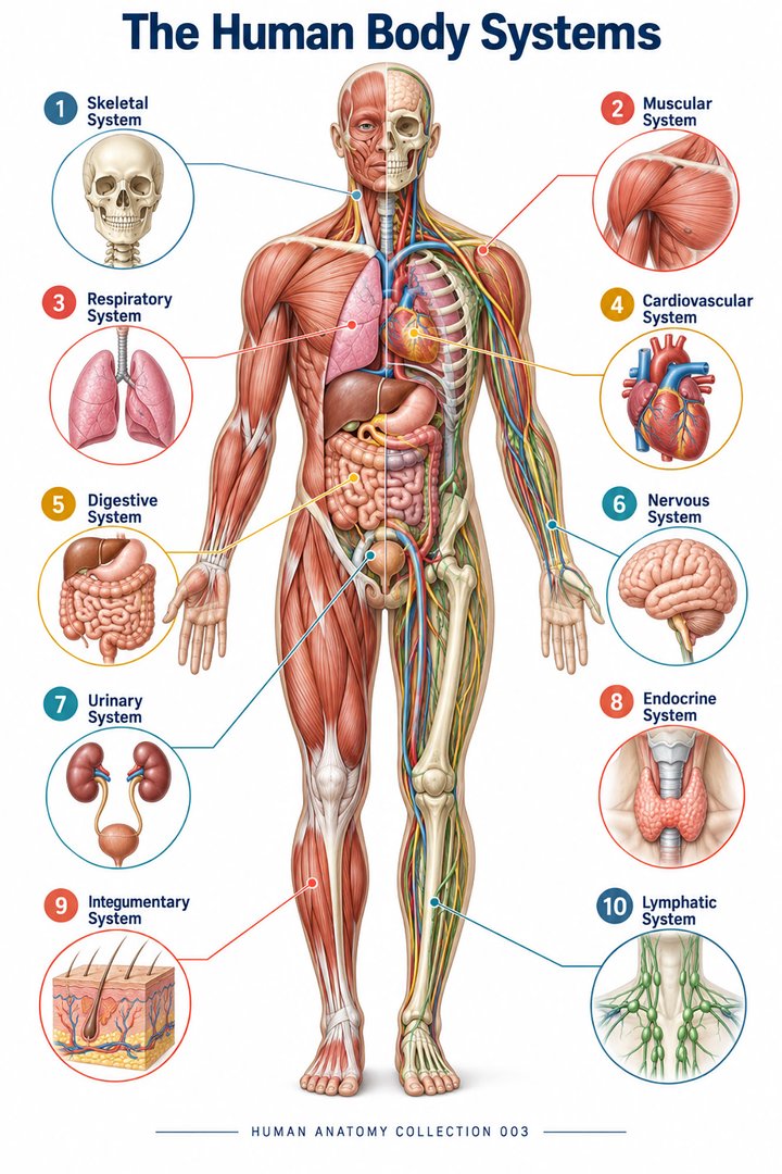 Human Body Systems ↗
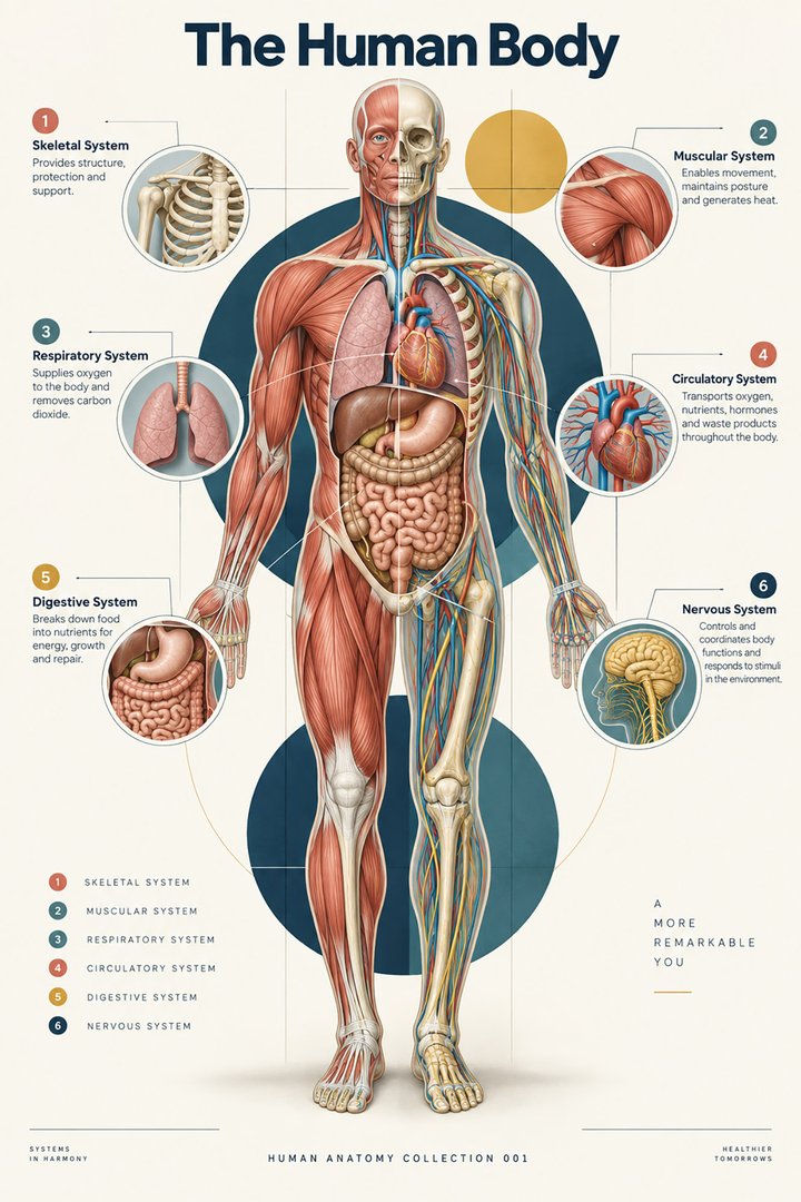 Human Body Facts ↗Human Anatomy and Physiology ↗
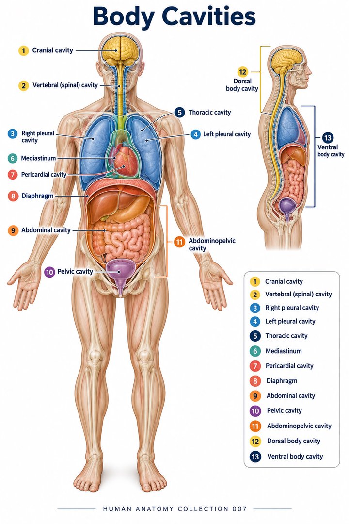 Human Body Cavities ↗Human Torso Anatomy ↗Human Digestive Tract ↗
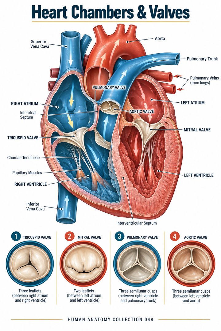 Human Heart Chambers ↗Human Organ Placement ↗
 Human Body Proportions ↗
 Peripheral Nervous System ↗
 Human Endocrine Glands ↗
 Human Bone Density ↗Human Blood Circulation ↗
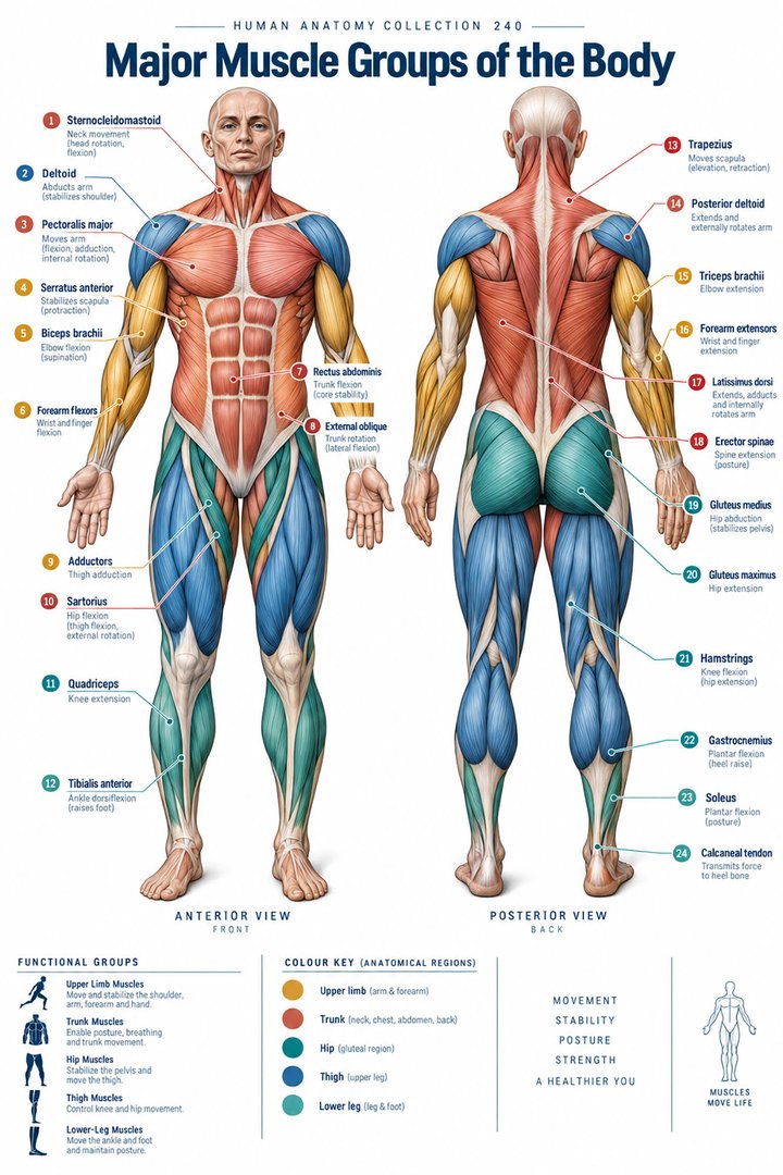 Human Muscle Groups ↗
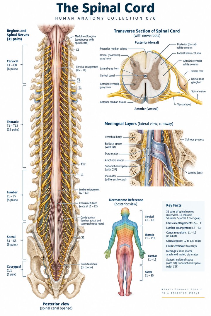 Human Spinal Cord ↗
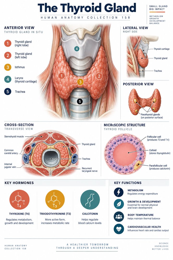 Human Thyroid Gland ↗
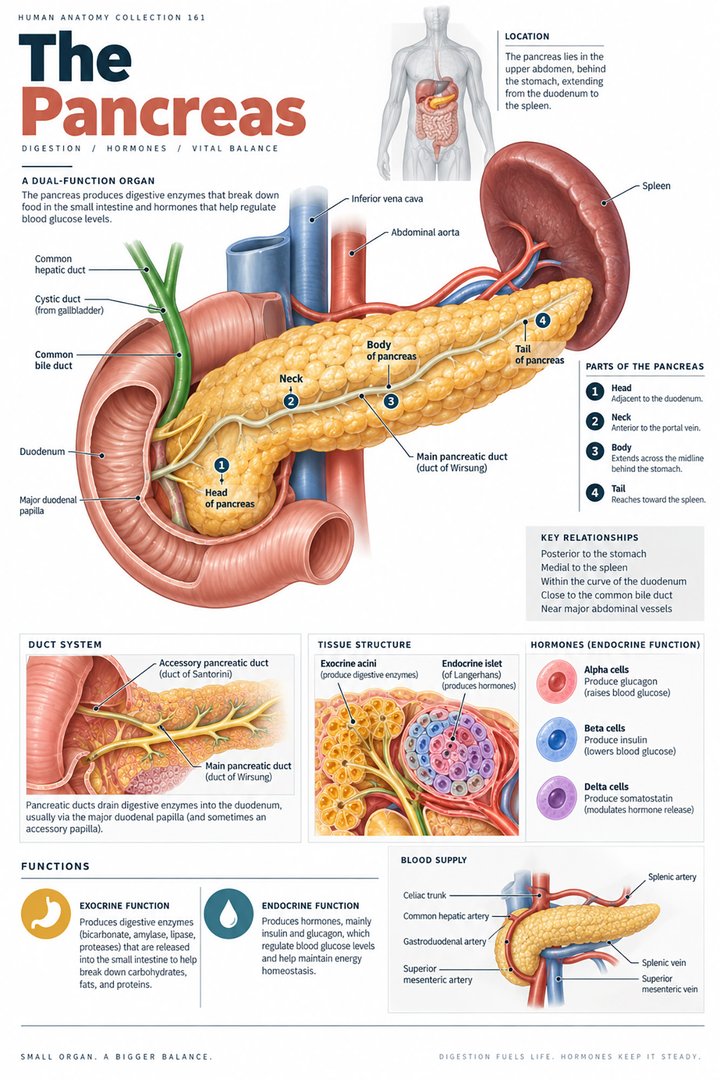 Human Pancreas Function ↗
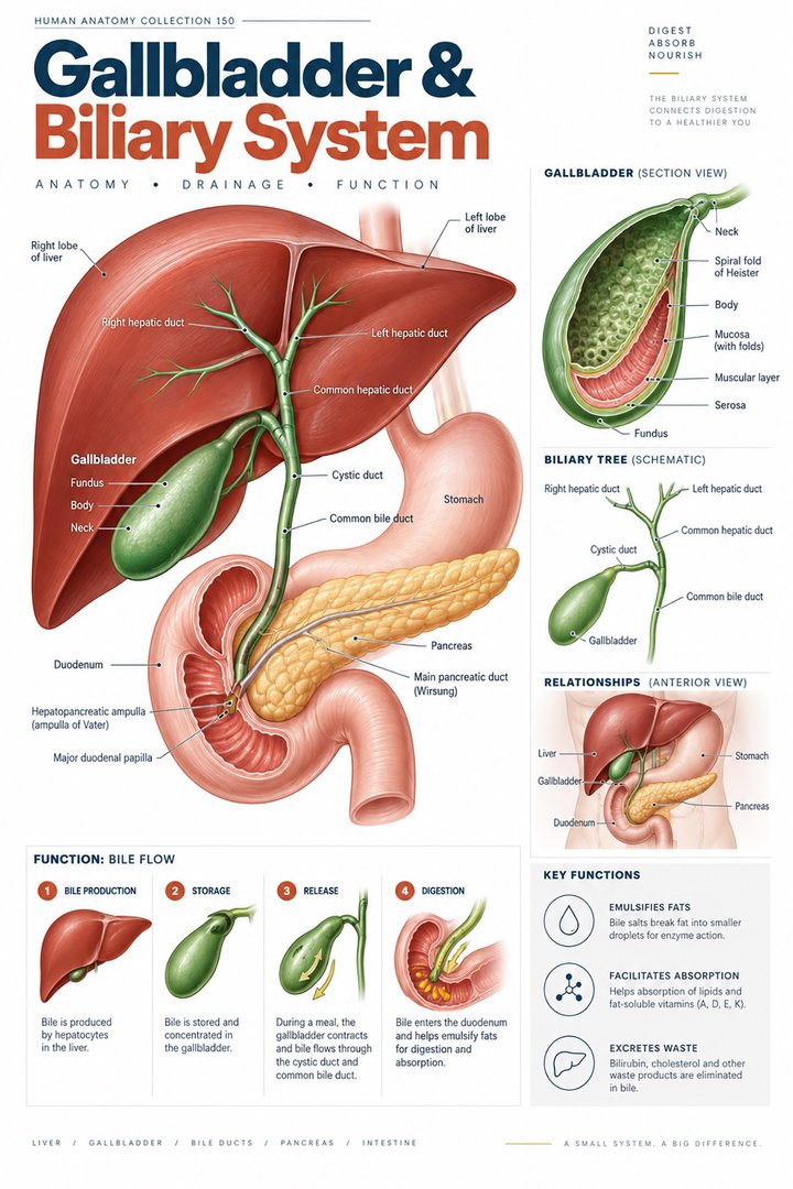 Human Gallbladder Anatomy ↗